- Author Rachel Wainwright [email protected].
- Public 2023-12-15 07:39.
- Last modified 2025-11-02 20:14.
Atrial septal defect
The content of the article:
- Causes and risk factors
- Forms of the disease
- Disease stages
- Symptoms
- Diagnostics
- Treatment
- Possible complications and consequences
- Forecast
Atrial septal defect (ASD) is a congenital defect, which is a non-closure (of different area) of the inner wall of the heart, which separates the right atrium from the left.

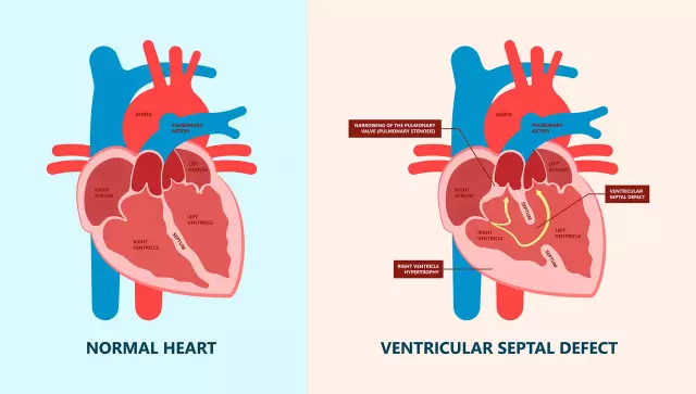
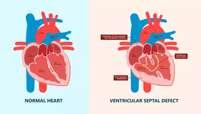
Picture of atrial septal defect
Normal hemodynamics implies the isolation of the parts of the heart: if normally the blood from the atrium moves freely into the corresponding ventricle, then the communication of the left and right atria or ventricles with each other is a pathology. The isolation of the heart sections ensures the immiscibility of oxygen-rich blood from the left sections and venous blood carrying carbon dioxide, or the blood of the "right heart".
In the prenatal period, the atrial septum (MPP) has an opening - the so-called oval window, which is necessary for normal blood circulation of the fetus. Since the function of external respiration is not realized at this stage, oxygen is delivered to the fetus through the umbilical vein. Having passed through the vasculature, blood from the umbilical vein enters the right atrium, from where a larger volume enters the open oval window and then into the left atrium, providing oxygen and nutrients to the brain tissue, the heart and the entire upper half of the trunk, and the upper limbs.
After the birth of a child, the blood flow in the lungs in connection with the beginning of the full functioning of the respiratory system becomes many times (up to 10) more intense, which leads to an increase in venous return and an increase in pressure in the left atrium. Changes in hemodynamics lead to the closure of the venous window with a valve from the left atrium - so the blood flows of the large and small circle are isolated and begin to function fully in the extrauterine period.
If the oval window does not close or there is another defect of the interatrial septum in a newborn, hemodynamics is impaired, structural elements of the heart are damaged, and the blood circulation of organs and tissues suffers.
Atrial septal defect is the most common congenital heart disease, accounting for approximately 1/5 of the total structure of congenital anomalies of the cardiovascular system. More common in girls.
Causes and risk factors
ASD substrate is a violation of the formation of the septum or the oval window valve in the embryonic period, which may be the result of several reasons:
- a genetic abnormality, often of a hereditary nature;
- the mother's use of prohibited substances, alcohol, as well as smoking;
- treatment with psychotropic, hormonal, antiepileptic, anti-tuberculosis, some antibacterial drugs in early pregnancy;
- exposure to ionizing radiation;
- professional harm to the expectant mother;
- viral diseases transferred by the mother in the first trimester of pregnancy (rubella, influenza);
- endocrinopathy.

Atrial septal defect is formed in the embryonic period under the influence of various factors
An open oval window may persist for some time after birth; the terms of spontaneous resolution of the anomaly are called different. It is more often indicated that the closure of the interatrial septal defect in children in half of the cases occurs before 1 year (according to other sources - up to 2 years). An atrial septal defect in adults (functioning oval window) in 25-30% of cases does not significantly affect hemodynamics.
The hemodynamic changes characteristic of this defect consist in the discharge of arterial blood from the left to the right atrium, which provokes an increase in blood volume in the pulmonary circulation and the development of pulmonary hypertension. Pulmonary hypertension in ASD leads to decompensation of the right heart.
Forms of the disease
Depending on the causative factor, there are:
- primary ASD (due to underdevelopment of the primary septum);
- secondary ASD (pathology of the formation of a secondary septum in embryogenesis);
- common atrium.
Secondary atrial septal defect is the most common and constitutes the bulk of MPP abnormalities. Depending on the localization, it can be of the following types:
- central defect;
- lower;
- upper;
- front;
- rear.
There is also a form with multiple defects.

Variants of atrial septal defects
Primary ASD is the rarest type of defect; they practically do not occur in isolation. In this case, the defect is usually located in the lower part of the septum directly above the atrioventricular opening.
Disease stages
Depending on the degree of stress of compensatory capabilities, stages of compensation and decompensation are distinguished.
Symptoms
An early sign of the possible presence of an atrial septal defect in children is pallor or cyanosis of the nasolabial triangle, which appears from the first days of life with crying, straining. There are no other specific signs in the early stages.

Cyanosis of the nasolabial triangle in a newborn is the first sign of an atrial septal defect
With a compensated defect, there are no pronounced symptoms. Indirect signs of ASD in this situation:
- lag in physical development;
- low body weight;
- frequent colds;
- rapid fatigue, poor exercise tolerance.
As a rule, by the age of 15-20, the compensatory capabilities of the cardiovascular system are depleted, there are clear signs of decompensation of the disease, active symptoms:
- shortness of breath on exertion, and in severe cases - at rest;
- palpitations;
- a feeling of interruptions in the work of the heart;
- cyanoticity, pallor of the skin and visible mucous membranes;
- cough on the background of intense physical activity, sometimes with sputum streaked with blood.
Diagnostics
Revealing the defect is not difficult. An atrial septal defect in a newborn is diagnosed during a planned mandatory ultrasound of the heart at the age of 1-1.5 months.
If the presence of a septal anomaly is confirmed, a second study is prescribed at the age of 1 year, based on the results of which a decision is made on the need for treatment.

The diagnosis of "atrial septal defect" is made on the basis of compulsory ultrasound of the heart at the age of 1-1.5 months
In addition to ultrasound of the heart, the presence of ASD is confirmed by the results of the following studies:
- auscultation (systolic murmur);
- phonocardiography (accent and splitting of the II tone, mesodiastolic or presystolic murmur over the tricuspid valve);
- chest x-ray [enlargement of the right heart, expansion and pulsation of the pulmonary artery and its branches ("dance of the roots"), congestion in the lungs];
- ECG [deviation of the electrical axis of the heart to the right, the specific configuration of the P waves (P-pulmonale)];
- coronary angiography (transition of contrast agent from the left to the right atrium during systole).
Treatment
The defect is treated with surgery.
Defect elimination is possible in several ways:
- open heart surgery in conditions of artificial circulation;
- endoscopic surgery;
- endovascular technique of defect closure with the introduction of an occluder through a catheter in the femoral vein.

Endovascular technique for closure of the atrial septal defect
Possible complications and consequences
Complications of an atrial septal defect can be:
- heart failure;
- recurrent pneumonia;
- septic endocarditis.
Forecast
With the timely detection of the anomaly and its surgical correction, the prognosis is favorable.
The prognosis is not determined until the child reaches the age of 1-2 years, since at this age the defect is able to resolve on its own. The prognosis worsens with late detection of the defect in adulthood, active signs of hemodynamic disorders, a large defect area, and the presence of concomitant diseases.
An atrial septal defect accidentally discovered during a routine examination in adults is not prognostically unfavorable and does not require surgery if it does not affect the quality of life and the patient has no complaints from the cardiovascular and respiratory systems.
YouTube video related to the article:

Olesya Smolnyakova Therapy, clinical pharmacology and pharmacotherapy About the author
Education: higher, 2004 (GOU VPO "Kursk State Medical University"), specialty "General Medicine", qualification "Doctor". 2008-2012 - Postgraduate student of the Department of Clinical Pharmacology, KSMU, Candidate of Medical Sciences (2013, specialty "Pharmacology, Clinical Pharmacology"). 2014-2015 - professional retraining, specialty "Management in education", FSBEI HPE "KSU".
The information is generalized and provided for informational purposes only. At the first sign of illness, see your doctor. Self-medication is hazardous to health!


