- Author Rachel Wainwright [email protected].
- Public 2023-12-15 07:39.
- Last modified 2025-11-02 20:14.
Astrocytoma
The content of the article:
- Causes and risk factors
- Forms of the disease
- Symptoms
- Diagnostics
- Treatment
- Possible complications and consequences
- Forecast
- Prevention
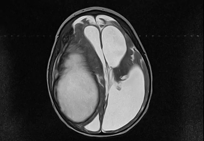
Astrocytoma is a primary brain tumor originating from the astrocytes (stellate cells) of the neuroglia. Astrocytomas differ in clinical course, degree of malignancy, and localization. The incidence is 5 to 7 cases per 100,000 population.
Astrocytomas of the brain can occur in people of any age and gender, but men 20-50 years old are more susceptible to them.
In adults, astrocytomas are usually localized in the white matter of the cerebral hemispheres. In children, they are more likely to affect the brainstem, cerebellum, or optic nerve.

Astrocytoma - glial brain tumor arising from astrocytes
Causes and risk factors
The exact causes leading to the development of astrocytomas are currently unknown. Contributing factors can be:
- viruses with a high degree of oncogenicity;
- hereditary predisposition to tumor diseases;
- some genetic diseases (lumpy sclerosis, Recklinghausen's disease);
- certain occupational hazards (rubber production, oil refining, radiation, heavy metal salts).
Forms of the disease
Depending on the cellular composition, astrocytomas are divided into several types:
- Pilocytic astrocytoma. Benign tumor (grade I), usually localized in the optic nerve, brainstem, cerebellum. It usually occurs in children. It is characterized by slow growth and clear boundaries.
- Fibrillar astrocytoma. According to the histological structure, it belongs to benign tumors, but has a tendency to recurrence (II degree of malignancy). Differs in slow growth and lack of clear boundaries. It does not grow into the meninges, does not give metastases. Fibrillar astrocytoma occurs in young people under 30 years of age.
- Anaplastic astrocytoma. Malignant tumor (III degree of malignancy), characterized by the absence of clear boundaries and rapid infiltrative growth. Most often affects men over 30 years old.
- Glioblastoma. Malignant and most dangerous type of astrocytoma (IV degree of malignancy). It has no boundaries, quickly grows into surrounding tissues and gives metastases. Usually seen in men between 40 and 70 years old.
Highly differentiated (benign) astrocytomas make up 10% of the total number of brain tumors, 60% are anaplastic astrocytomas and gliomas.
Symptoms
All symptoms of cerebral astrocytoma can be divided into general and local (focal). The development of general symptoms is due to an increase in intracranial pressure due to compression of the brain tissue by a growing tumor. The first manifestations of the disease are usually of a non-specific general nature:
- persistent headaches;
- dizziness;
- nausea, vomiting;
- lack of appetite;
- visual disturbances (the appearance of fog before the eyes, diplopia);
- increased lability of the nervous system;
- memory impairment, decreased performance;
- epileptic seizures.
The rate of progression of astrocytoma symptoms depends on the degree of malignancy of the tumor. But over time, focal symptoms join the general ones. Their appearance is associated with compression or destruction of the adjacent cerebral structures by a growing tumor. Focal symptoms are determined by the localization of the tumor process.

Focal symptoms of astrocytomas
When astrocytoma is located in the cerebral hemispheres, hemihypesthesia (impaired sensitivity) and hemiparesis (muscle weakness) of the extremities of one side of the body, opposite to the localization of the tumor, occur.
When a tumor of the cerebellum is damaged, coordination of movement is impaired, it becomes difficult for the patient to maintain balance while standing and when walking.
Astrocytomas of the frontal lobe of the brain are characterized by:
- decreased intelligence;
- memory impairment;
- attacks of aggression and pronounced mental agitation;
- decreased motivation, apathy, inertia;
- severe general weakness.
Astrocytomas localized in the temporal lobe of the brain are accompanied by hallucinations (gustatory, auditory, olfactory), memory impairment and speech disorder. Astrocytomas at the border of the occipital and temporal lobes can cause visual hallucinations.
The defeat of the tumor in the occipital lobe causes visual impairment.
With parietal astrocytoma, there are violations of fine motor skills of the hand, disorders of writing.
Diagnostics
If a brain astrocytoma is suspected, a clinical examination of the patient is carried out by a neurosurgeon, ophthalmologist, neurologist, otolaryngologist, psychiatrist. It should include:
- neurological examination;
- research of mental status;
- ophthalmoscopy;
- determination of visual fields;
- determination of visual acuity;
- examination of the vestibular apparatus;
- threshold audiometry.

Angiography is performed to diagnose astrocytoma and its features.
The primary instrumental examination in case of suspected astrocytoma of the brain consists in conducting electroencephalography (EEG) and echoencephalography (EchoEG). The identified changes are an indication for referral to magnetic resonance imaging or computed tomography of the brain.
To clarify the features of the blood supply to the astrocytoma, angiography is performed.
An accurate diagnosis with the definition of the degree of malignancy of the tumor can only be made based on the results of histological analysis. It is possible to obtain biological material for this study with stereotaxic biopsy or during surgery.
Treatment
The choice of a method for treating astrocytoma of the brain largely depends on the degree of its malignancy.
Removal of benign astrocytomas of small size (no more than 3 cm) is usually performed using stereotaxic radiosurgery. It provides targeted irradiation of tumor tissue with minimal radiation exposure to healthy tissue.

Brain tumor radiosurgery
Most astrocytomas are removed by conventional craniotomy surgery. In many cases, radical removal of malignant tumors is impossible, since they quickly grow into the surrounding brain tissue. To improve the patient's condition and prolong his life, surgeons resort to palliative operations aimed at reducing the volume of tumor formation and reducing the severity of hydrocephalus.
Radiation therapy for cerebral astrocytomas is usually given in inoperable cases. The course consists of 10-30 radiation sessions. In some cases, radiation therapy is prescribed in preparation for surgery because it can reduce the size of the tumor.

Radiation therapy for inoperable astrocytomas
Another method used for astrocytomas is chemotherapy. It is most commonly used to treat children. In addition, chemotherapy is prescribed in the postoperative period, because it reduces the risk of metastasis and tumor recurrence.
Possible complications and consequences
Astrocytomas of the brain, even benign ones, have a pronounced negative effect on the brain structures, causing a violation of their functions. The tumor can cause loss of vision, paralysis, mental disorders, etc. The progression of astrocytoma leads to compression of the brain and death.
Forecast
The prognosis is poor, due to the high degree of malignancy of most diagnosed tumors. In addition, benign astrocytomas can recur and become malignant. With astrocytomas of the brain of III and IV degrees of malignancy, the average life of the patient is one year. The most optimistic prognosis for astrocytomas of the 1st degree of malignancy, however, in this case, life expectancy in most cases does not exceed five years.
Prevention
Currently, there are no preventive measures aimed at preventing astrocytomas, since the exact reasons for their development have not been established.
YouTube video related to the article:

Elena Minkina Doctor anesthesiologist-resuscitator About the author
Education: graduated from the Tashkent State Medical Institute, specializing in general medicine in 1991. Repeatedly passed refresher courses.
Work experience: anesthesiologist-resuscitator of the city maternity complex, resuscitator of the hemodialysis department.
The information is generalized and provided for informational purposes only. At the first sign of illness, see your doctor. Self-medication is hazardous to health!


