- Author Rachel Wainwright wainwright@abchealthonline.com.
- Public 2023-12-15 07:39.
- Last modified 2025-11-02 20:14.
Lung tomography

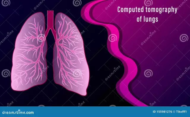
Lung tomography is one of the ways to diagnose lung diseases. It is carried out using a ring-shaped X-ray that takes pictures of the lungs from different angles. Computed tomography has become widespread, which makes it possible to digitize the images obtained, to provide numerous sections of the lungs for analysis.
Tomography of the lungs can be done in two modes: to assess the condition of the lungs and mediastinal organs: trachea, heart, superior vena cava, lymph nodes, pulmonary aorta and artery.
In some cases, when difficulties arise with the diagnosis, the patient is injected with a contrast in order to better see the lungs.
With the help of tomography, it is possible to identify chronic embolism, tuberculosis, diffuse diseases, lung cancer (localization of tumors, condition of lymph nodes, presence and prevalence of metastases), pneumonia, occupational diseases caused by inhalation of silicon particles, asbestos, quartz, etc.
Indications for the appointment of tomography
To make a tomography of the lungs is prescribed in such conditions and suspicion of them:
- changes in the structure of lung tissue;
- disorders in the thymus gland,
- fluid in the pleura;
- inflammation of the lymph nodes in the chest;
- diseases of the sternum and ribs;
- changes in the pericardium;
- neoplasms of benign and malignant nature in the lungs and pleura
- foreign bodies in the lungs;
- bronchiectasis
Preparation for tomography of the lungs
When prescribing such an examination, patients need to warn the doctor about pregnancy, breastfeeding, heart disease, asthma, thyroid problems, a history of myeloma, and claustrophobia.

Tomography of the lungs is a painless and short-term procedure, but a patient with increased emotional excitability and anxiety caused by the planned examination is allowed to take a sedative.
In some cases, anesthesia is practiced before the tomography, in order to ensure the immobility of the patient: children and "severe" patients.
Immediately before the procedure, the patient should undress to the waist, remove jewelry and items containing metal, and lie on the table. You cannot move during the examination. The presence of pacemakers is not a contraindication for tomography.
The patient is alone in the X-ray room, and the technologist who monitors the equipment is in the next room. Communication between them occurs through intercom.
The tomography results are ready immediately after the end of the examination and the radiologist can voice the main diagnosis to the patient. A detailed written opinion can be collected only after 1-2 days.
Can CT scan of the lungs of children be done?
Considering that tomography involves irradiation of the patient, when prescribing tomography of the lungs for children, not all parents consider it appropriate and are afraid of the serious consequences of exposure to x-rays.
When prescribing tomography of the lungs for children, the doctor proceeds from the considerations that the dose of radiation is small, the procedure itself lasts only 1-7 minutes, and cannot be dangerous, and the benefits of a timely diagnosis are many times greater than the estimated harm from radiation. Therefore, such an examination is allowed to be prescribed even to newborn children.
An alternative to lung tomography and radiation can be magnetic resonance imaging - a study based on the ability of hydrogen to emit electromagnetic waves. The possibility of using this method depends on the specifics of the disease and should be discussed with the attending physician.
Found a mistake in the text? Select it and press Ctrl + Enter.


