- Author Rachel Wainwright [email protected].
- Public 2024-01-15 19:51.
- Last modified 2025-11-02 20:14.
Left ovarian cyst
The content of the article:
- Varieties of left ovarian cysts
-
Functional cavity formations of the ovaries
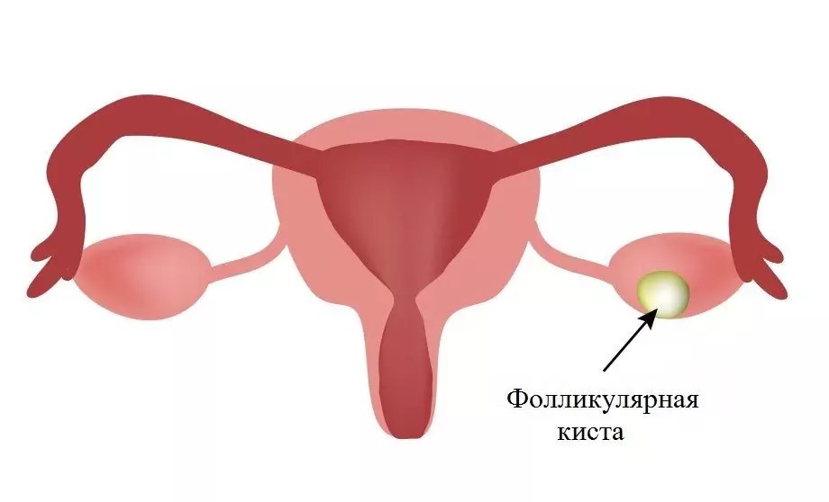
- Follicular ovarian cyst
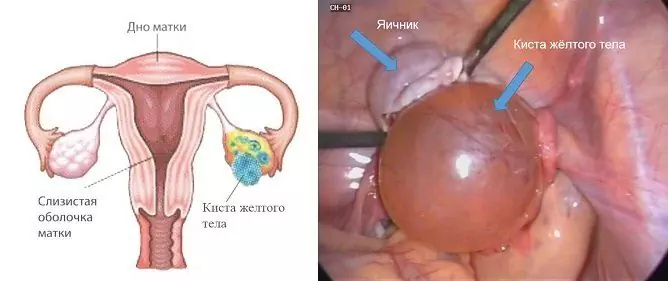
- Corpus luteum cyst
-
Non-functional cavity formations
- Endometrioma
- Paraovarian cyst
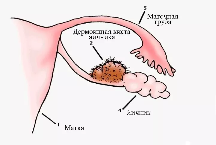
- Dermoid
- Left ovarian cyst symptoms
- Diagnostics
-
Left ovarian cyst treatment
- Wait-and-see tactics
- Treatment tactics depending on the type of cystic formation
- Video
The cyst of the left ovary, like that of the right, is a benign cavity neoplasm, consisting of an outer membrane and liquid internal contents. It is formed in the structure of the ovary and increases its volume due to the delay or excessive accumulation of fluid in the pre-existing cavity. In this it differs from true tumors of the gonads, the growth of which occurs due to cell proliferation. It occurs in women at any age. In the absence of complications, it is often asymptomatic. Therapeutic tactics are determined by the type and size of education, the presence and severity of clinical manifestations.

The cyst of the left ovary has the same causes of formation as the right
Varieties of left ovarian cysts
The most common types of ovarian cysts are:
- follicular;
- corpus luteum;
- paraovarian;
- endometrioid;
- dermoid.
The first two types are formed from the natural structures of the female reproductive glands - the follicle and the corpus luteum. They are called functional and respond to monthly cyclical fluctuations in hormone levels.
Functional cavity formations of the ovaries
The immediate cause of the appearance of functional cavity formations in the ovary is the lack of timely regression of the dominant follicle or corpus luteum.
Follicular ovarian cyst
A follicular cyst is the result of follicle persistence, a process by which the maturing dominant follicle does not ovulate in the middle of the menstrual cycle, but continues to grow. The accumulating follicular fluid stretches the follicular cavity, and a cystic formation appears. A formal sign of the transition of a follicle into a cyst is its size more than 3 cm.
Such cystic formations are one-sided, formed with the same frequency in both the left and right ovaries. They are round, single-chambered with a thin elastic capsule up to 8-10 cm in diameter, more often 5-6 cm. They usually appear after puberty, which confirms their hormonal dependence. The reason for the appearance is associated with a reduced hormonal function of the gonads, leading to an increase in the level of gonadotropic hormones of the anterior pituitary gland.
Corpus luteum cyst
The development of luteal, or corpus luteum cysts, is due to the fact that after ovulation, the follicular cavity does not collapse and is not completely filled with luteal cells, as it should be, but continues to exist and is stretched by serous fluid. The emerging cystic formation differs from the normal corpus luteum only in its large size (up to 7-8 cm in diameter).
Its walls are represented by luteal cells, which go through all stages of development of a normal corpus luteum:
- proliferation;
- vascularization;
- flourishing;
- reverse development.
Thus, the luteal cyst is a functioning cystic yellow body. Its formation is associated with increased production of gonadotropic hormones, it is also possible to develop against the background of the inflammatory process of the ovaries.
Non-functional cavity formations
Non-functional cavity formations of the female genital glands do not respond to monthly cyclical hormonal changes. They can originate from ovarian tissue, such as the endometrioma, or be of non-ovarian origin, such as the paraovarian cyst.
Endometrioma
Endometriosis is a disease in which cells from the inner lining of the uterus, or endometrium, enter other organs. Since the endometrial tissue has receptors for sex hormones, at any localization it is subject to cyclical changes, which are completely analogous to the processes occurring with the mucous membrane inside the uterus.
Minor bleeding from the focus of endometriosis that occurs monthly leads to the formation of a cavity filled with blood in the ovary. The latter, accumulating over time, thickens, darkens and becomes similar in consistency and color to liquid chocolate mass. This allows such cysts to be called "chocolate".
The etiology of endometriomas has not been finally established, several theories are being considered:
- implantation;
- embryonic;
- immune;
- migration.
There is no unified concept of the onset of pathology, but the presence of provoking factors is not disputed, including:
- hormonal disorders;
- inflammatory processes;
- heredity;
- abnormal position of the uterus.
Endometriomas can be both superficial and small, and large single, reaching 10-15 cm in diameter.
Attention! Photo of shocking content.
Click on the link to view.
Paraovarian cyst
In women, in the wide uterine ligament between the ovary and the tube, there is a peri-ovarian appendage, or paraophoron. It is represented by a network of thin small tubules, united into one channel, and is an organ that has lost its significance. When the secret in the lumen of the tubules is delayed and accumulates, an epididymal cyst is formed, which is called paraovarian.
The appendage develops as much as possible during the period of formation and flowering of the menstrual function. It is at this age that the cysts of the periobital appendage are most often detected. They are round or oval in shape with a smooth surface and transparent, watery contents. Sizes vary from 1-2 to 15-20 centimeters.
Education is located between the leaves of the broad ligament of the uterus. As it grows towards the abdominal cavity, one of the sheets protrudes with the formation of a leg, which includes the fallopian tube, and sometimes its own ovarian ligament.
Dermoid
The dermoid, or dermoid cyst, is a benign tumor - a mature teratoma that has external signs of a cyst and develops in violation of the processes of embryonic development. Sometimes the appearance of a dermoid is associated with trauma.
The dermoid usually forms on one side. It is grayish-whitish in color with a smooth surface, has great mobility due to its long stem, which creates favorable conditions for its twisting. The growth of the dermoid is slow, usually it does not reach large sizes.
The consistency of the dermoid is often uneven: elastic in some areas, dense to stony in others. The content is thick and resembles lard; hair, bones, teeth, rudiments of eyes, ears are often found in it.
Left ovarian cyst symptoms
Cystic formations of the gonads can be completely asymptomatic and detected during routine preventive examinations or ultrasound examination of the pelvic organs for another reason. The appearance of complaints is usually associated with large sizes and complications in the form of:
- rupture of the capsule;
- twisting of the legs;
- suppuration.
In this case, complaints come to the fore, the nature of which indicates a catastrophe in the abdominal cavity. Their list is as follows:
- intense cramping pains;
- nausea;
- vomiting;
- dizziness;
- weakness;
- attacks of fear;
- chills;
- pressure drop;
- decrease or complete cessation of intestinal peristalsis;
- increased body temperature.
A follicular cyst may be accompanied by a delay in menstruation. Endometriomas provoke various painful sensations that intensify on the eve and during menstruation, lead to a pronounced adhesive process in the small pelvis and cause infertility.
Large in diameter cavity formations squeeze the organs adjacent to the ovary. When it is the urinary tract, then women are worried about dysuric disorders: frequent urination, false desires, a feeling of incomplete emptying of the bladder. If this is the rectum, then bloating of the intestines, discomfort during bowel movements, constipation, or, conversely, increased stool frequency, are possible.
Diagnostics
A mass of the gonads can be detected by a gynecologist during examination, but the diagnosis is made after an ultrasound scan. Each type of cavity formations has characteristic echoes.
| View | Typical echoes |
| Follicular | Single-chamber formations of a round or oval shape up to 10 cm in diameter, the outline is even, clear, the wall is thin, no more than 2 mm, the contents are anechoic with acoustic amplification behind, along the periphery there is normal ovarian tissue. |
| Corpus luteum | A rounded formation with a thick wall, with CDC (color Doppler mapping) - a "ring of fire" along the periphery. In case of hemorrhage into the cavity, hyperechoic inclusions (fine suspension, a mesh of fibrin threads) are visualized, there is no blood flow inside. |
| Endometrioma | Rounded hypoechoic cavity formation with a double contour, wall thickness sometimes reaches 8 mm, the capsule may contain separate hyperechoic foci. The structure of the contents of the cavity is fine-celled, the shape of the cells is elongated or round, they can fill only a part of the volume. There are no dense inclusions and vessels in the lumen. |
| Paraovarial | An anechoic thin-walled formation, enclosed between the leaves of the broad ligament of the uterus, usually less than 5 cm in size. Above the cyst is the fallopian tube, next to the normal ovary. It is often possible to use a sensor to separate the cyst from the gonads. |
| Dermoid | A rounded hypoechoic formation with clear contours, single or multiple inclusions, behind them an acoustic shadow is visible, with CDK there is no vascularization. |

The main method for diagnosing ovarian cysts is ultrasound
If the cavity formation on ultrasound contains dense parietal structures, then to exclude oncological pathology, magnetic resonance imaging is performed, the level of tumor markers in the blood is determined: CA-125, HE-4.
Left ovarian cyst treatment
Wait-and-see tactics
Expectant tactics are used for functional neoplasms that respond to monthly fluctuations in hormones. Usually follicular and luteal formations exist for 2-3 cycles and dissolve on their own. Lack of reverse development and significant size require active action.
Expectant tactics are unacceptable with the development of complications: rupture of the cyst, twisting the legs, suppuration. The appearance of signs of an acute abdomen requires urgent specialized care.
Treatment tactics depending on the type of cystic formation
The task of the operation, carried out by the method of laparoscopy, is to remove the cystic formation with maximum preservation of healthy ovarian tissue. To prevent their recurrence, combined oral contraceptives are prescribed for a period of 3 to 6 months.
A corpus luteum cyst is often diagnosed during pregnancy. Since it is a functioning cystic yellow body, it usually disappears on its own by 12-16 weeks. Very rarely, they resort to removing it with rapid growth and significant size.
Small-sized paraovarial cavities can be observed; with growth and large volumes, they can be treated surgically. The formation is exfoliated from the intraligamentary space, and then the leaf of the wide uterine ligament is sutured, while the ovary and fallopian tubes are preserved.
Dermoid treatment is surgical. Rare recurrence and malignant transformation of the dermoid allow resection of the gonads with maximum preservation of microscopically unchanged tissue.
Patients with endometriomas are shown combined treatment - resection of the ovaries within healthy tissues and mandatory hormonal treatment. With surgery, if possible, restore normal anatomical relationships in the small pelvis. Removal of the endometrioma is carried out as carefully as possible, since its opening can lead to seeding of the peritoneum and the further development of the pathological process.
Video
We offer for viewing a video on the topic of the article.

Anna Kozlova Medical journalist About the author
Education: Rostov State Medical University, specialty "General Medicine".
Found a mistake in the text? Select it and press Ctrl + Enter.


