- Author Rachel Wainwright wainwright@abchealthonline.com.
- Public 2023-12-15 07:39.
- Last modified 2025-11-02 20:14.
Femur

The femur (Latin osfemoris) is the largest and longest tubular bone of the human skeleton, which serves as a lever of movement. Its body has a cylindrical shape somewhat curved and twisted along the axis, widened downward. The anterior surface of the femur is smooth, the posterior surface is rough, serving as the site of muscle attachment. It is subdivided into the lateral and medial lips, which are close to the middle of the femur adjacent to each other, and diverge downward and upward.
The lateral lip downwardly thickens and expands, passing into the gluteal tuberosity - the place to which the gluteus maximus muscle is attached. The medial lip descends below, turning into a rough line. At the very bottom of the femur, the lips gradually move away, limiting the popliteal surface of a triangular shape.
The distal (lower) end of the femur is somewhat widened and forms two rounded and fairly large condyles, differing from each other in size and degree of curvature. Relative to each other, they are located at the same level: each of them is separated from its "brother" by a deep intercondylar fossa. The articular surfaces of the condyles form a concave patellar surface, to which the patella is attached with its back side.
Femoral head
The head of the femur rests on the superior proximal epiphysis, connecting with the rest of the bone with the help of a neck that is spaced from the axis of the femur body at an angle of 114-153 degrees. In women, due to the greater width of the pelvis, the angle of inclination of the femoral neck approaches a straight line.
At the boundaries of the transition of the neck to the body of the femur there are two powerful tubercles, which are called trochanters. The location of the greater trochanter is lateral; the trochanteric fossa is located on its median surface. The lesser trochanter is located below the neck, occupying a medial position relative to it. In front, both trochanters - both large and small - are connected by an intertrochanteric crest.
Femur fracture
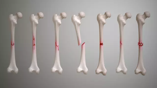
A femur fracture is a condition characterized by a violation of its anatomical integrity. Most often, it occurs in the elderly, when falling on its side. Concomitant factors of hip fractures in these cases are decreased muscle tone, as well as osteoporosis.
Signs of a fracture are severe pain, swelling, impaired function and limb deformity. Trochanteric fractures are characterized by more intense pain that worsens when trying to move and feel. The main symptom of a fracture of the upper part (neck) of the hip is the "sticky heel symptom" - a condition in which the patient cannot turn the leg at a right angle.
Femur fractures are divided into:
- Extra-articular, which, in turn, are divided into impacted (abduction), not impacted (adduction), trochanteric (intertrochanteric and pertrochanteric);
- Intra-articular, which include a fracture of the femoral head and a fracture of the femoral neck.
In addition, the following types of intra-articular hip fractures are distinguished in traumatology:
- Capital. In this case, the fracture line affects the femoral head;
- Subcapital. The fracture site is located immediately under its head;
- Transcervical (trancervical). The fracture line is in the area of the femoral neck;
- Basiscervical, in which the fracture site is located on the border of the neck and body of the femur.
If the fractures are punctured, when a fragment of the femur is wedged into another bone, conservative treatment is practiced: the patient is placed on a bed with a wooden shield placed under the mattress, while the injured leg rests on the Beller splint. Further, skeletal traction is carried out for the condyles of the lower leg and thigh.
In the case of displaced fractures, characterized by deformation and a vicious position of the limb, surgery is recommended.
Femur necrosis

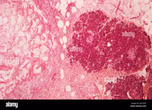
Femur necrosis is a serious disease that develops as a result of a violation of the structure, nutrition or fatty degeneration of bone tissue. The main cause of the pathological process that develops in the structure of the femur is a violation of blood microcirculation, osteogenesis processes and, as a result, the death of bone tissue cells.
There are 4 stages of femoral necrosis:
- Stage I is characterized by periodic pain radiating to the groin area. At this stage, the cancellous substance of the femoral head is damaged;
- Stage II is characterized by severe constant pain that does not disappear at rest. Radiographically, the head of the femur is speckled with small, eggshell cracks;
- Stage III is accompanied by atrophy of the gluteal muscles and thigh muscles, there is a displacement of the gluteal fold, shortening of the lower limb. Structural changes are about 30-50%, a person is prone to lameness and uses a cane for movement.
- Stage IV - the time when the femoral head is completely destroyed, which leads to the patient's disability.
The occurrence of femoral necrosis is facilitated by:
- Injuries of the hip joint (especially with a fracture of the femoral head);
- Household injuries and cumulative overloads, received during sports or physical activity;
- The toxic effects of certain drugs;
- Stress, alcohol abuse;
- Congenital dislocation (dysplasia) of the hip;
- Bone diseases such as osteoporosis, osteopenia, systemic lupus erythematosus, rheumatoid arthritis;
- Inflammatory, colds, which are accompanied by endothelial dysfunction.
The method of treatment of femoral necrosis depends on the stage of the disease, its nature, age and individual characteristics of the patient. To date, there are no drugs that can fully restore blood circulation in the femoral head, therefore organ restoration is most often performed by surgical methods. These include:
- Decompression of the femur - drilling several channels in the head of the femur, inside which vessels begin to form and grow;
- Fibula graft transplant;
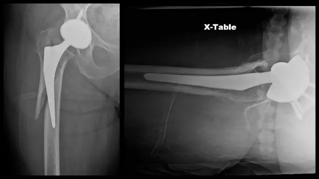
- Endoprosthetics, in which a damaged joint is replaced by a mechanical structure.
Found a mistake in the text? Select it and press Ctrl + Enter.

