- Author Rachel Wainwright wainwright@abchealthonline.com.
- Public 2023-12-15 07:39.
- Last modified 2025-11-02 20:14.
Spine
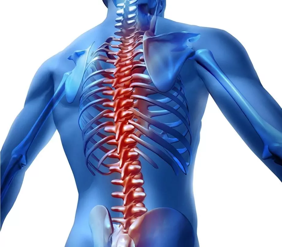
The spine is the main part of the axial skeleton of the body. The structure of the spine is ideally suited to support the function. In addition, the spine is a complex receptacle of the spinal cord and nerve roots.

The vertebral column is formed by 24 vertebrae and intervertebral discs. Intervertebral discs are biological shock absorbers that damp the loads constantly perceived by the spine, connect the vertebrae with each other, and also play the role of semi-joints, providing a small range of motion within one segment.
Spine sections
There are five sections of the spine:
- cervical (has 7 vertebrae);
- chest (12 vertebrae);
- lumbar (5 vertebrae);
- sacral (5 vertebrae);
- coccygeal spine (3-5 vertebrae).
Spine bends
The vertebral column has bends: lordosis - sections curved forward (cervical, lumbar), kyphosis - backward sections (thoracic, sacral).
Vertebra structure
Vertebrae are the bones that form the vertebral column. The anterior cylindrical portion of each vertebra is called the body. The vertebral body withstands the main axial load. Behind the body is a semi-circular arch of the vertebra, which has several processes. The vertebral body and arch limit the vertebral foramen. The openings of all vertebrae located one above the other form the spinal canal. The spinal canal contains the spinal cord, nerve roots, blood vessels, and fatty tissue.
7 processes extend from the arch of each vertebra: spinous process (unpaired) and paired upper articular, lower articular and transverse processes. The transverse and spinous processes provide attachment of ligaments and muscles, and the articular processes take part in the formation of the facet joints of the spine.
Spine ligaments
In addition to the bodies and arches of the vertebrae, ligaments are involved in the formation of the spinal canal. An important role is played by the posterior longitudinal, as well as the yellow ligament. The posterior longitudinal ligament looks like a strand, connects the bodies of all vertebrae from behind. The yellow ligament of the spine connects the arches of the adjacent vertebrae. She got the name due to the yellow pigment included in the composition. In the event of damage to the intervertebral discs and joints, these ligaments compensate for the pathological mobility (instability) of the vertebrae, while hypertrophy of the ligaments occurs. As a result of this process, the lumen of the spinal canal decreases, then even small hernias and bone growths squeeze the spinal cord and roots.
Intervertebral disc
An intervertebral disc is a flat, rounded formation located between adjacent vertebrae. The structure of the intervertebral disc is rather complex. In the center of the disc is the nucleus pulposus, which has elastic properties and acts as a shock absorber for the vertical load. Around the nucleus pulposus there is a multilayer annulus fibrosus, which holds the nucleus pulposus in the center, and also prevents the displacement of the vertebrae. The intervertebral disc of an adult has no vessels; its cartilage is nourished by the diffusion of oxygen and nutrients from the vessels of the vertebral bodies. This is why most drugs do not penetrate the disc cartilage. This complicates the treatment of the spine in many diseases.
Intervertebral joints
Intervertebral, or facet, joints connect adjacent vertebrae and are symmetrically located on both sides of the vertebral arch. The articular processes of adjacent vertebrae are directed towards each other, the ends of the processes are covered with articular cartilage. The cartilage has a slippery smooth surface, so that friction between the bones that form the joint is significantly reduced. The ends of the articular processes are surrounded by a sealed articular capsule. The cells of the joint capsule produce synovial fluid. It is essential for nourishing and lubricating the articular cartilage.
Intervertebral foramen
Intervertebral foramina are located in the lateral parts of the spine, formed by bodies, legs, and also articular processes of adjacent vertebrae. Veins and nerves exit through the intervertebral foramen, and arteries that supply the structures of the spine enter. There are two holes between a pair of vertebrae.
Spinal hernia
A spinal hernia, or intervertebral hernia, is a displacement of the nucleus pulposus located in the intervertebral disc, with rupture of the annulus fibrosus. Most often, this pathology occurs in the lumbosacral region, much less often in the cervical, thoracic regions. Clinical signs of a hernia of the spine are local pain in the projection of the damaged disc, which increases with exertion, radiating to the buttock, as well as along the back of the thigh, numbness, tingling in the innervation zone of the damaged roots, impaired sensitivity in the lower extremities, and dysfunction of the pelvic organs. With the localization of a hernia in the cervical spine, pain radiating to the upper limb, dizziness, arterial hypertension, numbness of the fingers are characteristic. When the thoracic region is affected, constant pain occurs when working in a forced position, localized in the thoracic spine. Treatment of herniated discs consists of systemic anti-inflammatory therapy (NSAIDs), as well as surgical treatment. Indications for surgical treatment of the spine in this pathology are neurological disorders and pain syndrome, resistant to conservative therapy. Surgical treatment consists of intralaminar microsurgical removal of the disc herniation. The direction of endoscopic removal of intervertebral hernia is also actively developing. Indications for surgical treatment of the spine in this pathology are neurological disorders and pain syndrome, resistant to conservative therapy. Surgical treatment consists of intralaminar microsurgical removal of the disc herniation. The direction of endoscopic removal of intervertebral hernia is also actively developing. Indications for surgical treatment of the spine in this pathology are neurological disorders and pain syndrome, resistant to conservative therapy. Surgical treatment consists of intralaminar microsurgical removal of the disc herniation. The direction of endoscopic removal of intervertebral hernia is also actively developing.
Spine fracture
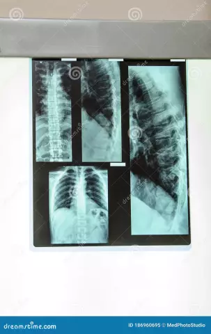
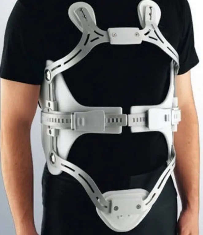
A spinal fracture is a violation of the anatomical integrity of the bones of the spine under the influence of force, provoking sharp excessive flexion movements in the spinal column, or directly under the influence of a traumatic agent. According to the mechanism of damage, such types of spinal fractures are distinguished: compression, flexion-distraction, rotational spinal fractures. The symptoms of a spinal fracture depend on the location of the injury. Typical symptoms are pain, numbness, muscle spasms, changes in intestinal motility, paralysis (this symptom indicates damage to the spinal cord). The diagnosis is made on the basis of X-ray examination data, computed tomography, magnetic resonance imaging of the spine. Treatment of a spinal fracture can be conservative (special retaining braces) or operative (necessary for displaced spinal fractures). In the treatment of compression fractures, vertebroplasty and kyphoplasty are used.
Found a mistake in the text? Select it and press Ctrl + Enter.


