- Author Rachel Wainwright wainwright@abchealthonline.com.
- Public 2023-12-15 07:39.
- Last modified 2025-11-02 20:14.
Liver cyst
The content of the article:
- What is a neoplasm
- Causes of liver cysts and their types
- Symptoms
- Why is a liver cyst dangerous?
- Diagnostics
-
Treatment
- Drug therapy
- Diet therapy
- Surgery
- Video
A liver cyst is a benign growth that is a cavity filled with fluid. According to statistics, cystic formations in the liver are recorded in 0.8-2% of the population. Pathology is more often found in adult women (30-50 years old).

Hepatic cysts may be an incidental diagnostic finding
What is a neoplasm
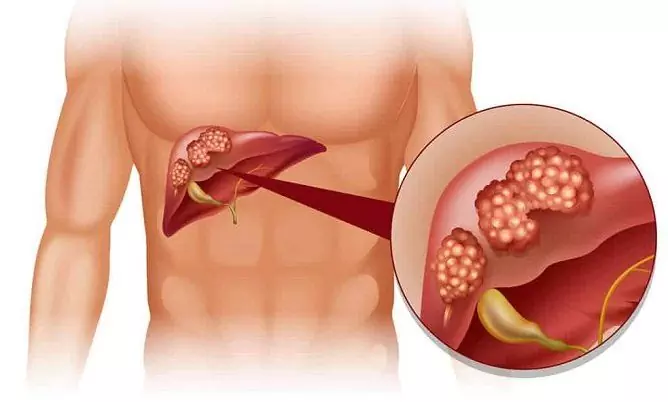
In the photo, the cyst is represented by a focal cavity neoplasm, which is filled with liquid contents and lined with a cylindrical or cubic epithelium. There are also so-called false cysts, their difference from the true ones is that they do not have their own wall - their wall becomes altered liver tissue.
Usually, the contents of the cystic cavity are transparent, colorless, in more rare cases, the neoplasm is filled with a liquid or jelly-like mass, which can have a brownish and / or greenish tint. With hemorrhage into a cyst, the contents become hemorrhagic, with the development of an infectious process - purulent.
Cystic formations can occur in various segments and lobes of the organ and reach large sizes (25 cm in diameter and more). As a rule, cysts of the left lobe of the liver develop more often. The cystic cavity can be localized on the surface or inside the organ, that is, its location can be subcapsular or parenchymal (intraparenchymal).
Causes of liver cysts and their types
Cystic formations can be congenital and acquired, false and true, single and multiple, as well as parasitic and nonparasitic.
False cystic formations often develop against the background of injuries, an inflammatory process, and may appear after surgical treatment of a liver abscess or other disease.
The appearance of parasitic cysts is a consequence of infection with parasites (helminthic invasion), mainly echinococcus.
True cysts are formations that arise during the prenatal period of development. This group includes:
| View | Explanation |
| Solitary | Single neoplasm |
| Plural | Cysts of the right lobe of the liver or left, occupy no more than 30% of the tissue, tissue between neoplasms is preserved |
| Polycystic | Neoplasms are localized in both lobes, occupy at least 60% of the entire tissue, there is no liver tissue between the walls of the formations |
| Cystofibrosis | In the presence of neoplasms of this type, there is an excessive proliferation of connective tissue in the organ, which displaces normal tissue |
The use of certain drugs (for example, drugs with estrogens, hormonal contraceptives), a history of infectious diseases can contribute to the development of cystic formation.
Symptoms
In the presence of small false cysts, obvious symptoms in a person are often absent, therefore, pathology is often detected during ultrasound examination (ultrasound) or computed tomography (CT) during diagnosis for another reason.
Symptoms usually occur when the cyst reaches 7-8 cm in diameter, as well as the presence of multiple formations that occupy more than 20% of the parenchyma volume.
In this case, the patient may experience:
- a feeling of heaviness and / or dull pain in the epigastric region, in the right side (may increase with walking, physical exertion);
- nausea and vomiting (usually after eating);
- decreased appetite;
- belching;
- flatulence;
- violation of defecation;
- weakness;
- excessive sweating;
- dyspnea;
- an increase in body temperature to subfebrile values;
- enlargement of the liver in size;
- jaundice;
- asymmetric enlargement of the abdomen;
- decrease in body weight.
This pathology can be combined with cystic formations of the bile ducts, cholelithiasis, polycystic kidney, pancreas and / or ovarian disease, cirrhosis, etc.
Why is a liver cyst dangerous?
The progression of the pathological process can lead to the development of a number of consequences dangerous for the liver: dysfunction, atrophy of organ tissues, replacement of the hepatic parenchyma with neoplasms.
Over time, polycystic disease can lead to the development of liver failure. Against the background of cystofibrosis, portal hypertension, hepatic failure, and cirrhosis of the liver often occur. Jaundice occurs when the bile ducts are compressed by an enlarging neoplasm.
Complications of a cyst can be:
- perforation;
- suppuration;
- rupture (entails hemorrhage and spread of infection);
- malignancy (degeneration into a malignant tumor).
With a hemorrhage, the patient usually has an attack of abdominal pain, and peritonitis may develop.
When the infection is attached, the liver may develop. If a person has echinococcal cystic formations, there is a risk of spreading the pathogen by the hematogenous route, while the patient may develop infectious foci in other organs, for example, in the lungs.
Diagnostics
For the diagnosis, ultrasound, CT / MRI, laboratory blood tests (liver function tests) are used.
In order to exclude the parasitic etiology of the neoplasm, a serological blood test (by enzyme immunoassay, indirect hemagglutination reactions) and a number of other laboratory tests can be carried out. In doubtful cases, diagnostic laparoscopy may be required.

Hepatic cysts can grow significantly in organ tissue
Differential diagnosis is carried out with tumors of the small intestine, pancreas, hemangioma, dropsy of the gallbladder, metastatic cancer.
Treatment
In the absence of any clinical signs, patients usually require dispensary observation by a gastroenterologist. If parasitic cysts are found, treatment is carried out under the supervision of a parasitologist (infectious disease specialist).
Drug therapy
Symptomatic therapy of pathology may consist in the use of analgesic, anti-inflammatory drugs, if necessary (infectious inflammation) - antibiotics, with parasitic cystic formations, anthelmintic drugs are prescribed.
Diet therapy
If the patient has cystic formation and / or after its removal, it may be necessary to follow a diet. Fried, fatty, salty, spicy, smoked food, canned food, carbonated drinks, strong coffee, sweets should be excluded from the diet. Patients are shown fractional meals (frequent eating in small portions). It is recommended to consume more foods rich in fiber, vitamins. The diet should include vegetables, fruits, berries, herbs, dairy products, fish.
After surgery to remove a cystic formation, the patient may need to follow a gentle diet throughout his life.
Surgery
To treat cystic formation with surgery is indicated in the following cases:
- compression of the portal vein system with the development of portal hypertension;
- the presence of severe symptoms that significantly impair the patient's quality of life;
- relapses after previous treatment;
- the risk of capsule rupture or other complications.
Surgical interventions that can be performed with cystic liver formations are of three types:
- Conditionally radical. Conditionally radical methods include excision of the walls of a cystic formation or its exfoliation (enucleation). Whenever possible, such operations are performed with a gentle laparoscopic approach.
- Radical. With solitary cystic formation, a radical method of treatment is liver resection; with polycystic disease, organ transplantation may be indicated.
- Palliative. In this case, the removal of the formation is not carried out. Puncture aspiration of the fluid contained in the cyst can be carried out, followed by the introduction of sclerosing drugs into the cavity. The neoplasm can also be opened, emptied and drained, etc. When the formation is localized in the gate of the liver, the cystic cavity can be emptied and its walls sutured to the edges of the surgical wound (marsupialization). In polycystic disease (in the absence of signs of hepatic and renal failure), fenestration can be performed, which is a partial excision of the walls of the formation.
In the postoperative period, it is required to avoid physical exertion, give up bad habits, and strengthen the immune system.
Video
We offer for viewing a video on the topic of the article.

Anna Aksenova Medical journalist About the author
Education: 2004-2007 "First Kiev Medical College" specialty "Laboratory Diagnostics".
The information is generalized and provided for informational purposes only. At the first sign of illness, see your doctor. Self-medication is hazardous to health!


