- Author Rachel Wainwright wainwright@abchealthonline.com.
- Public 2023-12-15 07:39.
- Last modified 2025-11-02 20:14.
Left ventricle
The left ventricle is one of the four chambers of the human heart, in which the systemic circulation begins, providing a continuous flow of blood in the body.

The structure and structure of the left ventricle
Being one of the chambers of the heart, the left ventricle in relation to other parts of the heart is located posteriorly, to the left and downward. Its outer edge is rounded and is called the pulmonary surface. The volume of the left ventricle in the course of life increases from 5.5-10 cm3 (in newborns) to 130-210 cm3 (by the age of 18-25).
Compared with the right ventricle, the left one has a more pronounced oblong-oval shape and is somewhat longer and more muscular.
In the structure of the left ventricle, two sections are distinguished:
- The posterior section, which is the cavity of the ventricle and with the help of the left venous opening, communicates with the cavity of the corresponding atrium;
- The anterior section - the arterial cone (in the form of an excretory canal) communicates with the arterial opening with the aorta.
Due to the myocardium, the wall of the left ventricle reaches 11-14 mm in thickness.
The inner surface of the wall of the left ventricle is covered with fleshy trabeculae (in the form of small protrusions), which form a network, intertwining with each other. Trabeculae are less pronounced than in the right ventricle.
Left ventricular function
The aorta of the left ventricle of the heart begins the systemic circulation, which includes all branches, the capillary network, as well as the veins of tissues and organs of the whole body and serves to deliver nutrients and oxygen.
Left ventricular dysfunction and treatment
Left ventricular systolic dysfunction is called a decrease in its ability to eject blood into the aorta from its cavity. This is the most common cause of heart failure. Systolic dysfunction is usually caused by a drop in contractility, leading to a decrease in its stroke volume.
Diastolic dysfunction of the left ventricle is called a drop in its ability to pump blood into its cavity from the pulmonary artery system (otherwise, to provide diastolic filling). Diastolic dysfunction can lead to the development of pulmonary secondary venous and arterial hypertension, which are manifested as:
- Cough;
- Dyspnea;
- Paroxysmal nocturnal dyspnea.
Pathological changes and treatment of the left ventricle
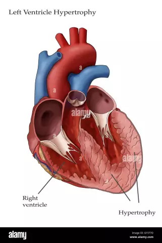
One of the typical heart lesions in hypertension is left ventricular hypertrophy (otherwise known as cardiomyopathy). The development of hypertrophy provokes changes in the left ventricle, which leads to a modification of the septum between the left and right ventricles and a loss of its elasticity.
Moreover, such changes in the left ventricle are not a disease, but represent one of the possible symptoms of the development of any type of heart disease.
The cause of the development of left ventricular hypertrophy can be both hypertension and other factors, for example, heart defects or significant and frequent exercise. The development of changes in the left ventricle is sometimes noted over the years.
Hypertrophy can provoke significant changes that occur in the region of the walls of the left ventricle. Along with the thickening of the wall, there is a thickening of the septum located between the ventricles.
Angina is one of the most common signs of left ventricular hypertrophy. As a result of the development of pathology, the muscle increases in size, atrial fibrillation occurs, and the following are also observed:
- Chest pain;
- High blood pressure;
- Headaches;
- Pressure instability;
- Sleep disturbances;
- Arrhythmia;
- Pain in the region of the heart;
- Poor health and general weakness.
In addition, such changes in the left ventricle can be symptoms of diseases such as:
- Pulmonary edema;
- Congenital heart disease;
- Myocardial infarction;
- Atherosclerosis;
- Heart failure;
- Acute glomerulonephritis.
Treatment of the left ventricle is most often of a medical nature, along with adherence to diet and rejection of existing bad habits. In some cases, surgery may be required to remove the portion of the heart muscle that has undergone hypertrophy.
Small heart anomalies, manifested by the presence of cords (accessory connective tissue muscle formations) in the cavity of the ventricles, is the false notochord of the left ventricle.
Unlike normal chords, left ventricular pseudochords have atypical attachment to the interventricular septum and free ventricular walls.
Most often, the presence of a false chord of the left ventricle does not affect the quality of life, but in the case of their multiplicity, as well as in an unfavorable location, they can cause:
- Serious rhythm disturbances;
- Decreased exercise tolerance;
- Relaxation disorders of the left ventricle.
In most cases, treatment of the left ventricle is not required, but should be monitored regularly by a cardiologist and prophylaxis of infective endocarditis.
Another common pathology is left ventricular heart failure, which is observed with diffuse glomerulonephritis and aortic defects, as well as against the background of the following diseases:
- Hypertonic disease;
- Atherosclerotic cardiosclerosis;
- Syphilitic aortitis with damage to the coronary vessels;
- Myocardial infarction.
Insufficiency of the left ventricle can manifest itself both in an acute form and in the form of a gradually increasing circulatory failure.
The main treatment for left ventricular heart failure is:
- Strict bed rest;
- Long-term inhalations with oxygen;
- The use of cardiovascular drugs - cordiamine, camphor, strophanthin, corazole, korglikon.
Found a mistake in the text? Select it and press Ctrl + Enter.


