- Author Rachel Wainwright wainwright@abchealthonline.com.
- Public 2023-12-15 07:39.
- Last modified 2025-11-02 20:14.
Hypertensive retinal angiopathy - what is it?
The content of the article:
- Risk factors for the development of retinal angiopathy in hypertensive type
- Signs of retinal angiopathy
- Treatment of hypertensive retinal angiopathy
- Video
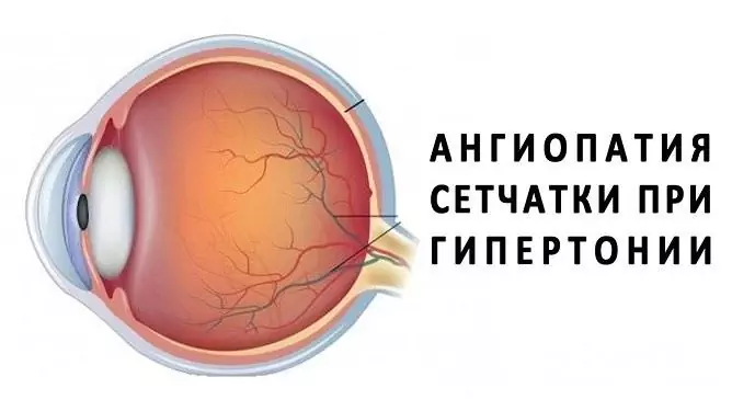
Hypertensive retinal angiopathy - what is it? This is a lesion of the blood vessels of the retina of the eye, caused by arterial hypertension (a persistent increase in blood pressure above 130 by 90 mm Hg).
Since retinal angiopathy of the hypertensive type is a complication of hypertension, there is no separate ICD-10 code for this pathological condition. The code of essential (primary) hypertension, against which this pathology develops, is I10. The code for peripheral angiopathy in diseases classified elsewhere is I79.2 *.

Retinal angiopathy is common, especially in advanced stages of hypertension
Retinal angiopathy is often diagnosed in women during pregnancy and in people over 35 years old. The most common hypertensive retinal angiopathy in both eyes. In some cases, pathology can lead to disability.
The diagnosis of hypertensive angiopathy is established on the basis of the definition of arterial hypertension in the patient and the associated pathological changes in the blood vessels of the retina of the eye and the optic nerve head.
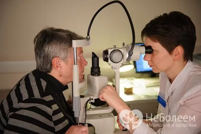
The study of the fundus is carried out with the obligatory dilation of the pupils. To determine the state of microcirculation, it may be necessary to conduct a contrast study of the blood vessels of the fundus.
Risk factors for the development of retinal angiopathy in hypertensive type
The development of arterial hypertension in humans can be caused by a genetic predisposition, endocrine diseases, pathologies of the cardiovascular system, kidneys, overweight, smoking, drinking alcoholic beverages, excessive consumption of table salt, a passive lifestyle, taking a number of medications, and frequent stressful situations. In some cases, the exact cause of an increase in blood pressure (BP) in a patient cannot be determined.
A prolonged increase in blood pressure causes changes in the structure of the vascular wall, disruption of the blood supply to organs and tissues, which, in turn, entails a disruption in their functioning. There is no clear connection between the features of the course of arterial hypertension in a patient and the severity of changes that occur with the blood vessels of the eye. Damage to the retina of the eye can develop both with hypertension of the 1st degree, and in the later stages of pathology. In this case, the individual features of the structure of the blood vessels of the eyes are of no small importance. Angiopathy is more often recorded in people with large arterial trunks of retinal vessels and straight fistulas.
Risk factors for the development of hypertensive retinal angiopathy are smoking, alcohol abuse, diabetes mellitus, autoimmune diseases, atherosclerosis, occupational hazards, overweight, a large amount of fast carbohydrates and animal fats in the diet. In addition, abnormalities in the structure of retinal vessels can contribute to pathology.
Signs of retinal angiopathy
With arterial hypertension, patients develop visual disturbances (blurred, black dots before the eyes), headache, dizziness, increased sweating, rapid pulse, apathy, drowsiness, irritability, facial swelling, numbness of the fingers. The clinical signs of angiopathy go through several stages, during which a number of changes occur with the formation of arterial deformities.
Functional changes (stage 1) are characterized by narrowing of the arteries and some dilatation of the veins, which leads to a slight disturbance of microcirculation, however, the changes that have occurred at this stage can only be determined by an ophthalmological examination with an examination of the fundus.
The further development of the pathology causes organic changes (stage 2) in the walls of the arteries (thickening, replacement by connective tissue). The hardening of the arteries causes a violation of the blood supply to the retina, blood outflow may be difficult due to compression of the veins by the arteries. This stage is characterized by a more pronounced violation of microcirculation. The patient has small areas of retinal edema, hemorrhage. On examination, the arteries appear narrowed, and the veins appear convoluted and dilated. The characteristic shine of the blood vessels is noted, which is caused by the compaction of the vascular wall.
With a critical violation of microcirculation, retinal functions are disrupted, and the disease passes into the stage of angioretinopathy, or stage 3. During the examination of the fundus, soft (areas of microinfarction) and hard (fatty deposits in the tissue) exudates are revealed. The arteries narrow even more, the swelling and the number of hemorrhages increase. With fatty deposits, yellowish spots appear on the eyes. If the lesion of the optic nerve joins, the patient develops neuroretinopathy, vision decreases, the likelihood of its complete loss increases significantly.
A patient with retinal angiopathy has intermittent blurred vision due to fluctuations in blood pressure. Visual acuity can remain normal even with pronounced organic changes in the vessels. It decreases in case of damage to the central region of the retina (in the event of edema), fatty deposits, hemorrhages, etc. With further progression of the disease, thrombosis of the central retinal vein or its branches, occlusion of the central retinal artery or its branches is possible.
Treatment of hypertensive retinal angiopathy
An ophthalmologist is involved in the treatment of the disease, however, if complications develop, other specialists may also take part in it.
First of all, a pressure correction is required. To normalize it, diuretic drugs, angiotensin-converting enzyme inhibitors, beta-blockers, calcium antagonists can be prescribed. Effective treatment of retinal angiopathy without correcting blood pressure is impossible.
With hemorrhages in the retina, drugs are prescribed that improve blood circulation and microcirculation, vasodilators, vitamins.
In the case of vascular complications, treatment is carried out in a hospital.
If there is a risk of retinal rupture or persistent hemorrhage, laser coagulation is indicated.

The diagnosis is established by the results of an ophthalmological examination.
Patients with hypertensive retinal angiopathy should limit the intake of fluids, sodium chloride, animal fats. The diet should include fermented milk products, lean meats and fish, vegetables and fruits.
Patients and people at risk should avoid excessive physical exertion, stress, give up bad habits, control body weight. Normalization of blood cholesterol levels is also important. Prolonged work at the computer and other activities associated with excessive eye strain are contraindicated.
Video
We offer for viewing a video on the topic of the article.

Anna Aksenova Medical journalist About the author
Education: 2004-2007 "First Kiev Medical College" specialty "Laboratory Diagnostics".
Found a mistake in the text? Select it and press Ctrl + Enter.


