- Author Rachel Wainwright wainwright@abchealthonline.com.
- Public 2024-01-15 19:51.
- Last modified 2025-11-02 20:14.
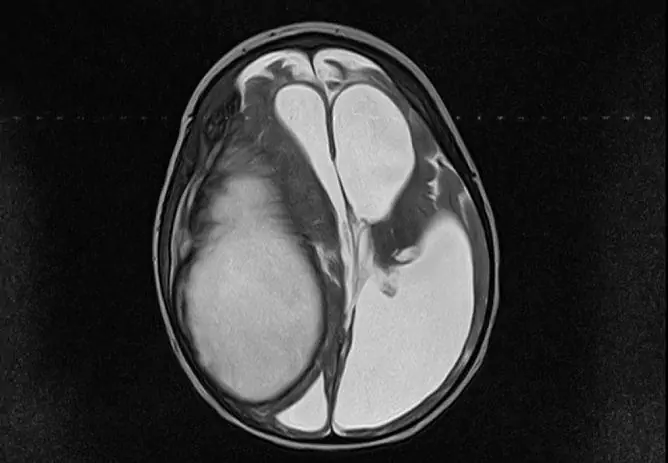
CSF cyst of the brain
The content of the article:
- Classification
-
The reasons
- Primary neoplasms
- Secondary neoplasms
- Clinical stages
- Symptoms
- Diagnostics
- Treatment
- Forecast
- Prevention
- Video
CSF cyst of the brain (arachnoid cyst) is a benign neoplasm located between the hard and arachnoid (arachnoid) membranes. At the site of formation, the arachnoid membrane is divided into two sheets and thickens. Cerebral fluid accumulates between these sheets, which leads to the development of a cystic cavity.

CSF, or arachnoid cyst is located between the arachnoid and hard membranes of the brain
According to medical statistics, the disease occurs in about 4-5% of adults. However, in most cases, CSF cysts are insignificant and do not manifest themselves clinically, so the data may turn out to be inaccurate. Only with an increase in the size of the neoplasm begin to put pressure on the brain, which causes the appearance of symptoms of intracranial (intracranial) mass formation.
Arachnoid cystic formations are most often localized above the Turkish saddle, in the area of the Sylvian groove or cerebellopontine angle.
Classification
By origin, cystic neoplasms are:
- congenital (primary) - arise even at the stage of intrauterine development;
- acquired (secondary) - pathological processes (bleeding, inflammation, traumatic injuries) occurring in the meninges become their causes.
Depending on the features of the morphological structure, cavity arachnoid formations are divided into two types:
- simple - their cavity is lined with cells that produce cerebral fluid;
- complex (retrocerebellar) - their structure includes various tissues (cells of the arachnoid membrane, neuroglia).
According to the characteristics of the clinical course, there are:
- frozen - they are characterized by a latent course, lack of growth;
- progressive - accompanied by a gradual increase in neurological symptoms, which is associated with an increase in the size of the formation and compression of the brain structures by it.
The reasons
Primary neoplasms
Congenital CSF cysts are associated with anomalies in the structure of the brain, which are formed at the stage of intrauterine formation of the organ. The factors that can provoke their development include negative effects on the body of a pregnant woman and the developing fetus:
- intoxication (taking medications with teratogenic effects, drug addiction, smoking, alcohol abuse, occupational hazards);
- infections (cytomegalovirus, herpes, rubella, toxoplasmosis);
- overheating (frequent baths, sunbathing);
- radiation exposure.
Secondary neoplasms
The causes of secondary CSF cysts are:
- craniocerebral trauma (bruises, concussions);
- neurosurgical interventions;
- meningitis;
- meningoencephalitis;
- arachnoiditis;
- subdural hematoma or subarachnoid hemorrhage.
Clinical stages
Clinically, CSF formations begin to manifest themselves only as their size increases. This is due to the fact that the volume of the skull is limited. The growing tumor begins to squeeze the structures of the brain, which leads to an increase in intracranial pressure, the appearance of neurological symptoms. In accordance with this, several stages of the clinical course of the disease are distinguished, which are presented in the table:
| Stage | Description |
| Clinical compensation | There are no signs of brain damage, despite the presence of a tumor in the cranial cavity |
| Clinical subcompensation | A person has the first signs of impaired brain function |
| Partial clinical decompensation | Characterized by persistent neurological damage |
| Gross clinical decompensation | There is a dislocation (displacement of structures), wedging into the foramen magnum. Against this background, the patient begins to show initial signs of a violation of basic vital functions. |
| Terminal | Disorders in the regulation of respiration and cardiovascular activity are growing, which ultimately becomes the cause of death |
Symptoms
Clinical manifestations of CSF cystic formation include cerebral and focal symptoms:
| Symptom group | Description |
| Cerebral |
Their development is associated with an increase in intracranial pressure. The patient develops persistent headaches that are bursting in nature. They usually intensify at night and early in the morning. At the height of the painful attack, nausea occurs, and there may be repeated vomiting. Mental disorders (psychomotor agitation, impaired critical abilities) may develop. When examining the fundus, congestion is detected. |
| Focal |
They are caused by local damage to certain brain structures. When the formation is localized in the region of the frontal lobes, the motor function suffers to a greater extent and the patient develops muscular hypertonicity, instability appears when walking, and speech disorders may occur. Lesions of the parietal lobe are characterized by impaired perception and sensitivity. Signs of damage to the temporal lobe are hallucinations, convulsive syndrome, defects in auditory perception. Localization of the cerebrospinal fluid cyst in the occipital lobes causes a dysfunction of the visual analyzer. With this form of the disease, the patient develops symptoms such as cortical blindness, visual agnosia, and visual illusions. |
Diagnostics
It is possible to establish the presence of an intracranial mass formation according to the data of a neurological examination and the results of an initial examination, which includes the following methods:
- echoencephalography (Echo-EG);
- rheoencephalography (Rheo-EG);
- electroencephalography (EEG).
In order to clarify the localization and nature of the volumetric neoplasm, MSCT and MRI of the brain with contrast are shown. The use of a contrast agent makes it possible to carry out differential diagnostics between the CSF cyst and another formation, since the contrast can accumulate in the liquid contents of the cystic cavity.

Based on the diagnostic results, a decision is made on the need for surgical treatment of cystic neoplasms.
Treatment
If the patient is diagnosed with a frozen CSF cyst with clinical compensation, then treatment is not indicated for him. In this case, the patient should be registered with a neurologist with a mandatory annual MRI scan.
With a progressive cyst in the stage of clinical subcompensation, the patient is prescribed conservative therapy aimed at lowering intracranial pressure. If it is ineffective, surgical intervention is indicated. The choice of the method of operation in each specific case is carried out by a neurosurgeon, taking into account the characteristics of the disease, the age of the patient, the presence or absence of concomitant pathology.
Currently, the following surgical techniques are most commonly used:
- Complete excision. The indication for surgery is a rupture of the capsule or hemorrhage into the cavity of the formation. The intervention is quite traumatic, and the recovery period is long.
- Endoscopic fenestration. A small hole is made in the bones of the skull with a cutter. Through it, puncture and aspiration of the liquid contents of the neoplasm are performed, followed by the creation of holes between its cavity and the subarachnoid space or ventricles of the brain. After that, the cavity in the arachnoid membrane is no longer filled with fluid and therefore the disease does not progress.
- Cystoperitoneal shunting. The essence of this operation is that a path is created for the outflow of cystic contents into the abdominal cavity, where it is absorbed into the blood.
Forecast
With a frozen CSF cyst, the prognosis is favorable. Such formations can exist throughout the patient's life without any symptoms. The danger is represented by progressive neoplasms. If diagnosed and treated late, they can cause persistent neurological deficits, patient disability and even death.
Prevention
Prevention of congenital CSF cysts is based on the prevention of congenital malformations and includes the following areas:
- exclusion of exposure to teratogenic factors on a pregnant woman;
- rational management of pregnancy;
- gentle delivery.
Prevention of acquired CSF cysts is based on the timely and complete treatment of vascular and inflammatory cerebral diseases, traumatic brain injuries.
Video
We offer for viewing a video on the topic of the article.

Elena Minkina Doctor anesthesiologist-resuscitator About the author
Education: graduated from the Tashkent State Medical Institute, specializing in general medicine in 1991. Repeatedly passed refresher courses.
Work experience: anesthesiologist-resuscitator of the city maternity complex, resuscitator of the hemodialysis department.
Found a mistake in the text? Select it and press Ctrl + Enter.


