- Author Rachel Wainwright wainwright@abchealthonline.com.
- Public 2023-12-15 07:39.
- Last modified 2025-11-02 20:14.
Sebaceous glands

Sebaceous glands are skin glands whose secret is a fatty lubricant for the surface of the skin and hair.
They are located almost all over the skin, with the exception of the soles of the feet and the skin of the palms. They differ significantly in size, and also have a different structure and localization in different areas of the skin. Most of the accumulation of sebaceous glands is observed in the scalp, as well as on the chin and cheeks. They are also located on areas devoid of hair: in the corners of the mouth, on the lips, clitoris, nipples, labia minora, glans penis, foreskin.
The location, size and structure of the sebaceous glands depends on the time the hair was laid. They are located in the reticular (reticular) layer of the dermis and lie in a slightly oblique direction between the hair lifter muscle and its follicle. When the muscle contracts, the hair straightens and applies pressure, promoting the secretion of the sebaceous gland.
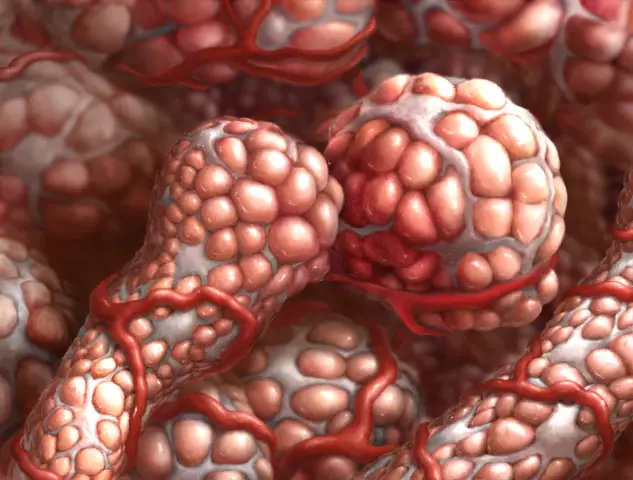
The simple sebaceous gland consists of an excretory duct and an end secretory part. The excretory duct is a non-keratinizing squamous epithelium lined from the inside, and the end secretory part is a pouch surrounded by a thin connecting capsule on the outside. Under the capsule, there is a growth layer - undifferentiated cells with high mitotic activity, which lie on the basement membrane. In the center of the end secretory part is cellular detritus, consisting of decayed secretory cells - the secret of the gland.
The blood vessels provide blood supply to the glands and nourish the hair root system. The sebaceous glands are supplied with adrenergic and cholinergic nerve fibers. Adrenergic nerve fibers pierce the basement membrane and surround secretory cells, while cholinergic nerve fibers are located on the surface of the basement membrane.
Throughout a person's life, the sebaceous glands change. At the time of birth, they are very developed and function intensively. During the first year of life, against the background of a reduced secretion of the sebaceous glands, their growth predominates, later they partially atrophy, especially in the skin of the back and legs.
During puberty, their growth increases again, the function of the sebaceous glands increases.
In old age, the glands stop developing, which is manifested in a decrease in their size, simplification of their structure, and a decrease in the functional and metabolic activity of secretory cells. Some glands disappear with age.
Normally, iron per day secretes about 20 g of sebum. The secretion of the sebaceous glands makes the hair elastic, regulates water evaporation, softens the epidermis, prevents the penetration of certain substances into the skin from the outside, and has an anti-fungal and antimicrobial effect.
The function of the sebaceous glands is regulated by the neurohumoral route, mainly by sex hormones, which can sometimes cause an increase in their activity (secretion of too much secretion, hyperplasia). In newborns, the glands are strongly influenced by the pituitary maternal hormones and progesterone, and during puberty they are affected by the activation of the gonadotropic function of the anterior pituitary gland, increased activity of the gonads, and activation of the adrenal cortex.
Sebaceous gland pathologies
Pathology of the glands consists of malformations, dystrophic changes, functional disorders, inflammation of the sebaceous glands and tumors of the glands.

Dysfunction of the sebaceous glands, as a rule, occurs as a result of damage to the autonomic peripheral or central nervous system, metabolic disorders and hormonal regulation. Often, an increase in the activity of the glands can be observed with catatonic stupor, lesions of the anterior lobe of the pituitary gland, gonads, adrenal cortex, as well as with epidemic viral encephalitis due to damage to autonomic centers. A decrease in the function of the glands leads to a decrease in the function of the endocrine glands, for example, during orchiectomy.
Often as a result of a malfunction, a blockage of the sebaceous glands occurs. One of the most common pathologies, which is based on a violation of the secretory function, is seborrhea. With this disease, sebaceous-horny plugs, or comedones, occur in the ducts of the glands, resulting from blockage of the sebaceous glands. Also, a frequent occurrence is the occurrence of atheromas - retention cysts. Multiple cysts also occur in pilosebocystomatosis due to a violation of nevoid dysplasia of the epidermis.
Dystrophic changes can be both age-related and develop with some acquired diseases (scleroderma, skin atrophy, etc.). Often dystrophic changes are caused by hereditary factors.
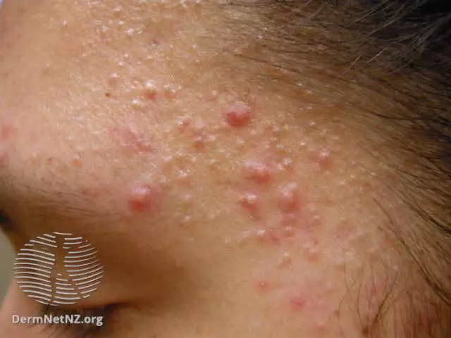
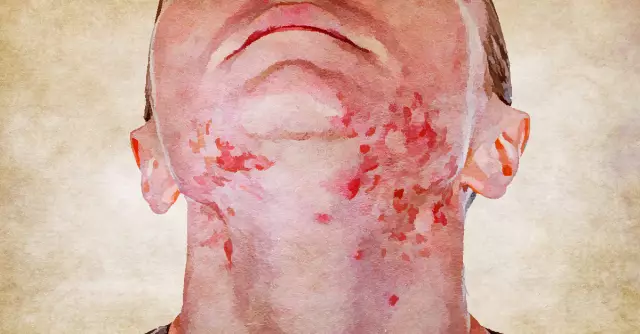
Inflammation of the sebaceous glands occurs quite often, especially during puberty. Inflammation is characterized by the formation of acne. In this case, the inflammatory process can capture the walls of the glands and tissues around them (pustular acne), and can spread into deeper layers of the skin, sometimes even capturing the subcutaneous tissue (phlegmous acne).
Benign tumors of the sebaceous glands include a true adenoma of the gland, which most often develops in adults and the elderly and looks like a dense round nodule on the back or face.
The basal cell carcinoma, which has a locally destructive growth, belongs to malignant tumors. Most often, cancer of the sebaceous gland develops from the glands of the cartilage of the eyelids - the meibomian glands.
Found a mistake in the text? Select it and press Ctrl + Enter.


