- Author Rachel Wainwright [email protected].
- Public 2023-12-15 07:39.
- Last modified 2025-11-02 20:14.
Mediastinum
Mediastinal structure

The mediastinum is the anatomical space, the median region of the chest. In front, the mediastinum is bounded by the sternum, and behind by the spine. On the sides of this organ are the pleural cavities.
For various purposes (surgical intervention, planning radiation therapy, describing the localization of pathology), the mediastinum, in accordance with the scheme proposed by Twining in 1938, is divided into upper and lower, as well as anterior, posterior and middle sections.
Anterior, middle, posterior mediastinum
The anterior mediastinum is bounded anteriorly by the sternum, posteriorly by the brachiocephalic veins, the pericardium and the brachiocephalic trunk. This space contains the internal thoracic veins, the thoracic artery, the mediastinal lymph nodes and the thymus - the thymus gland.
The structure of the middle mediastinum: heart, hollow veins, brachiocephalic veins and brachiocephalic trunk, aortic arch, ascending aorta, phrenic veins, main bronchi, trachea, pulmonary veins and arteries.
The posterior mediastinum is bounded by the trachea and pericardium in the anterior part, and in the posterior part by the spine. In this part of the organ are the esophagus, the descending aorta, the thoracic lymphatic duct, the semi-unpaired and azygous veins, as well as the posterior lymph nodes of the mediastinum.
Upper and lower mediastinum
All anatomical structures that lie above the upper edge of the pericardium belong to the superior mediastinum: its boundaries are the superior aperture of the sternum and a line drawn between the angle of the chest and the intervertebral disc Th4-Th5.
The inferior mediastinum is bounded by the upper edges of the diaphragm and pericardium and, in turn, is also divided into anterior, middle, and posterior parts.
Classification of neoplasms of the mediastinum
Organ neoplasms are considered not only true tumors of the mediastinum, but also tumor-like diseases and cysts that are different in etiology, localization and course of the disease. Each of the neoplasms of the mediastinum comes from tissues of different origins, united only by anatomical boundaries. They are classified into:
- Primary neoplasms;
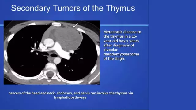
- Secondary malignant tumors (metastases of tumors of organs located in the lymph nodes of the mediastinum);
- Tumors of the organs entering the mediastinum;
- Tumors of tissues whose task is to restrict the mediastinum;
- Pseudo-neoplastic diseases (Benier-Beck-Schaumann disease, lesions of the mediastinal nodes in tuberculosis, parasitic cysts, malformations of large vessels, etc.).
The clinical picture of neoplasms

Mediastinal tumors are detected mainly in young and middle age with the same frequency, both in men and women. Despite the fact that mediastinal diseases may not manifest themselves for a long time and are detected only on a preventive study, there are several symptoms that characterize violations of this anatomical space:
- Intense pain localized at the site of neoplasms and radiating to the neck, shoulder, interscapular region;
- Dilatation of the pupil, drooping of the eyelid, retraction of the eyeball - can occur if a tumor grows into the borderline sympathetic trunk;
- Hoarseness of the voice - originates from the defeat of the recurrent laryngeal nerve;
- Severity, noise in the head, shortness of breath, chest pain, cyanosis and swelling of the face, swelling of the veins of the chest and neck;
- Violation of the passage of food through the esophagus.
In the late stages of mediastinal diseases, there is an increase in body temperature, general weakness, arthralgic syndrome, heart rhythm disturbances, edema of the extremities.
Mediastinal lymphadenopathy
Lymphadenopathy or enlargement of the lymph nodes of this organ is observed with metastases of carcinoma, lymphomas, as well as some non-neoplastic diseases (sarcoidosis, tuberculosis, etc.).
The main symptom of the disease is generalized or localized enlargement of the lymph nodes, however, mediastinal lymphadenopathy can have such additional manifestations as:
- Increased body temperature, sweating;
- Weight loss;
- Frequent infection of the upper respiratory tract (tonsillitis, pharyngitis, tonsillitis);
- Hepatomegaly and splenomegaly.
The defeat of the lymph nodes, characteristic of lymphomas, can be isolated or combine the invasion of tumors into other anatomical structures (trachea, blood vessels, bronchi, pleura, esophagus, lungs).
Found a mistake in the text? Select it and press Ctrl + Enter.


