- Author Rachel Wainwright wainwright@abchealthonline.com.
- Public 2023-12-15 07:39.
- Last modified 2025-11-02 20:14.
Panniculitis
The content of the article:
- Causes and risk factors
- Forms of the disease
- Symptoms
- Diagnostics
- Panniculitis treatment
- Possible complications and consequences
- Forecast
- Prevention
- Video
Panniculitis is an inflammation of adipose tissue, often subcutaneous, but sometimes localized elsewhere in the body, including the surrounding internal organs. The inflammatory process covers fatty lobules or interlobular septa.
Both adults and children are ill, women more often than men.

Panniculitis is more common in the limbs, but it can also affect the trunk.
Causes and risk factors
The mechanism of development of the disease is unknown. Causal factors can be:
- infections (streptococcal infection is the main cause of erythema nodosum, a form of panniculitis);
- systemic diseases (systemic sclerosis, vasculitis, systemic lupus erythematosus);
- intestinal inflammatory diseases;
- intoxication;
- sarcoidosis;
- histiocytosis;
- lymphoproliferative diseases;
- alpha-1-antitrypsin deficiency;
- pathologies of the pancreas, characterized by excessive activity of pancreatic enzymes;
- injuries (including operating ones), hypothermia.
Forms of the disease
Depending on the origin, the following forms of pathology are distinguished: infectious, enzymatic, traumatic, autoimmune, etc. When the cause cannot be identified, panniculitis is called idiopathic.
One of the most common forms of the disease is idiopathic lobular (nodular) panniculitis, which is called Weber-Christian disease. Synonyms: febrile recurrent non-suppressing panniculitis, chronic recurrent Weber-Christian panniculitis.
There are also nodal form (erythema nodosum), plaque, infiltrative, visceral (internal), mixed. One of the rare forms is mesenteric panniculitis - a chronic nonspecific inflammation of the adipose tissue of the intestinal mesentery, omentum and peritoneal tissue.
Symptoms
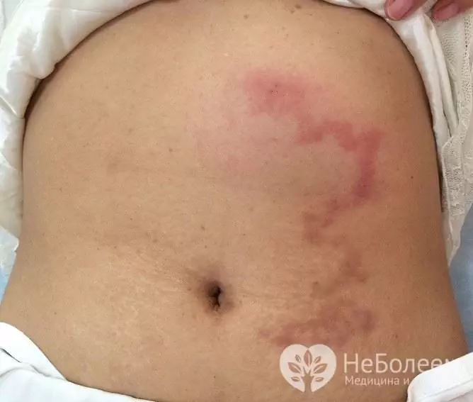
Panniculitis is manifested by the formation of subcutaneous erythematous nodes up to 5 cm in diameter, characterized by intense pain. They are usually located on the lower limbs, but can occur on the buttocks, back, abdomen, buttocks, chest, and face. Less commonly, in the scrotum, lungs, abdominal cavity (mesenteric panniculitis), and the skull.
The symptoms of different forms of panniculitis have some differences.
| The form | Manifestations |
| Plaque |
Individual nodes merge into plaques covered with purple or bluish skin. The plaques can be extensive, covering the entire surface of the limb. Against their background, lymphostasis often develops due to compression of blood vessels and nerve endings. |
| Infiltrative | In the area of the nodes, the subcutaneous fat melts, fistulas are formed, from which pus-like contents come out, which, unlike pus, are sterile, that is, it does not contain infectious pathogens. |
| Mixed | The transition from one form to another is characteristic - it begins with a nodular, which transforms into a plaque, and then into an infiltrative. |
The main symptom of Weber-Christian disease is the nodes located in the subcutaneous fatty tissue, usually located in the region of the limbs, less often on the trunk. The appearance of nodules is preceded by musculoskeletal pain and an increase in temperature to subfebrile values (no higher than 38 ° C). The nodules are sharply painful, the skin above them is changed, sharply hyperemic or purple-cyanotic. They are observed for several weeks, after which they heal, leaving behind rounded scars. Sometimes the nodes break through with the release of a yellowish oily discharge (infiltrative form), which subsequently also leads to scarring. Inflammation can be widespread with the involvement of joints, liver, kidneys, serous membranes, bone marrow.
Diagnostics
The diagnosis is made on the basis of a characteristic clinical picture, laboratory findings and radiography.
Laboratory diagnostics:
- clinical blood test - neutrophilia, increased ESR, anemia;
- biochemical blood test - increased serum lipase activity;
- clinical analysis of urine - erythrocytes, increased leukocyte count, proteinuria.
A biopsy of inflammatory elements is performed. In the samples taken, macrophages, necrotic fat cells, microvascular thrombosis, fibrosis at the site of tissue destruction are found.
If the joints are affected, radiography can reveal a decrease in joint spaces and foci of osteolysis.
Differential diagnosis is carried out with infectious diseases of the skin and subcutaneous adipose tissue.
Panniculitis treatment
No specific treatment has been developed; therapy is supportive. Patients are shown bed rest. To reduce pain, cold compresses are applied to the affected area, as well as non-steroidal anti-inflammatory drugs. With systemic damage, glucocorticosteroids are prescribed, in some cases, chemotherapeutic agents, immunosuppressive drugs are indicated.
Possible complications and consequences
A rare but severe complication of panniculitis is bone marrow involvement. Its functional impairment can be fatal.
Forecast
The prognosis is moderately favorable, it depends on the form of the disease, its cause, main and concomitant pathology.
Prevention
Since the mechanism of the disease is unclear, preventive measures have not been developed.
Video
We offer for viewing a video on the topic of the article.

Anna Kozlova Medical journalist About the author
Education: Rostov State Medical University, specialty "General Medicine".
The information is generalized and provided for informational purposes only. At the first sign of illness, see your doctor. Self-medication is hazardous to health!


