- Author Rachel Wainwright wainwright@abchealthonline.com.
- Public 2023-12-15 07:39.
- Last modified 2025-11-02 20:14.
Xanthelasma
The content of the article:
- Causes
- Forms of xanthelasm
- Signs of xanthelasm
- Diagnostics
- Xanthelasma treatment
- Prevention
- Consequences and complications
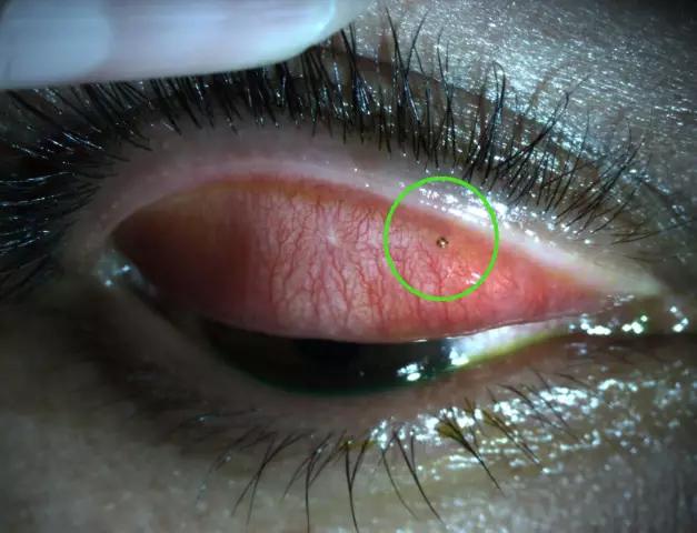
Xanthelasmas are single or multiple flat, round plaques that occur predominantly on the skin of the eyelids and are lipid deposits in the papillary dermis.

Source: glazexpert.ru
Xanthelasma is more common in females after 50 years. They belong to benign neoplasms, are histologically similar to xanthomas, however, unlike xanthomas, they are rarely accompanied by an increase in the concentration of triglycerides in the blood.
Xanthelasma of the eyelids can act as a marker of severe forms of atherosclerosis, in the same capacity it indicates an increased risk of developing myocardial infarction. Thus, the risk of developing coronary heart disease in patients with xanthelasma is 50%, heart attack (angina pectoris) - 40%, the risk of death from cardiovascular events in the presence of xanthelasma reaches 17% (adjusted for other risk factors).
Causes
The mechanism of the formation of xanthelasmas is not completely clear. Neoplasms develop in some patients against the background of impaired lipid metabolism. Most often they appear in overweight people suffering from diabetes mellitus, myxedema, pancreatitis, liver cirrhosis, lipoid nephrosis, cardiovascular diseases. They can also be hereditary (hereditary forms of lipid metabolism disorders).
Forms of xanthelasm
Xanthelasmas are single and multiple, and in some cases they take on a disseminated multiple character - in this case, they speak of skin xanthomatosis.
Signs of xanthelasm
Xanthelasmas appear suddenly, their formation is not preceded by any changes in the skin of the eyelid. They develop slowly, over months, and sometimes years, can reach the size of a large bean, have a straw-yellow or orange tint, a soft texture, a smooth (in some cases slightly wrinkled) surface, slightly rise above the skin.
Usually xanthelasmas appear on the skin of the upper eyelid. It can form on the lower eyelid, but the lower eyelid is rarely affected in isolation, usually in combination with the upper eyelid. Xanthelasmas are often combined with xanthomas on other parts of the body (especially on the elbows and knees), and can also be found on mucous membranes. Several xanthelasmas located nearby can merge into one, form lumpy elements, go on the bridge of the nose.
Sometimes, due to multiple xanthelasmas, the entire upper is crossed by a strip of irregular outlines. The appearance of neoplasms is not accompanied by any subjective sensations. They are not inclined to independent reverse development.
With primary hypercholesterolemic xanthomatosis, which belongs to hereditary liposes, xanthelasma can appear in children from the first year of life. In this case, they are combined with bone cysts, disorders of the cardiovascular system, liver.
Diagnostics
For the diagnosis of xanthelasma, an objective examination is usually sufficient, since the neoplasms have a characteristic appearance and localization. To reliably distinguish it from other neoplasms, the method of diascopy is used, which consists in examining the xanthelasma when pressing on it with a glass slide, which allows it to bleed and clearly determine the yellow color of the element.
Since xanthelasmas are a sign of impaired metabolism, an endocrinologist's consultation is necessary to diagnose the pathology that caused it.
Patients with xanthelasma are shown a biochemical blood test (lipid profile, determination of glucose concentration). The amount of additional research depends on the underlying disease, against which the pathological process has developed.
Differential diagnosis is carried out with malignant neoplasms of the skin, elastic pseudoxanthoma, syringoma.
Xanthelasma treatment
Treatment consists in treating the underlying disease that caused the appearance of xanthelasma. In order to correct lipid metabolism, drugs of lipotropic and hypocholesterolemic action are prescribed, if necessary, hormonal, hypoglycemic drugs. The main treatment can be supplemented by herbal medicine (birch buds, dandelion root, corn stigmas, immortelle flowers, plantain juice, rose hips, etc.).
An important condition for the normalization of lipid metabolism is a diet with limited use or exclusion from the diet of high-cholesterol foods, saturated fatty acids. Animal fats should be replaced with vegetable fats. The daily norm of butter should not exceed 25 g, and vegetable oil - 75 g. Fatty meats, offal (liver, kidney, brain), fatty cottage cheese, cheese, bakery products made from premium flour, coffee are excluded from the diet. It is recommended to limit the consumption of table salt to 3-4 g per day, as well as to stop drinking alcohol.
The diet should include low-starchy vegetables, fruits, berries, wholemeal bread, buckwheat, oats, brown rice, lentils, beans, fish, poultry, veal, beef, lamb, flaxseeds, nuts, milk and dairy products. Food should be steamed, boiled, stewed or baked. The recommended water regime is to consume at least 1.5 liters of water per day.
Xanthelasma is not dangerous and does not have the ability to degenerate into a malignant tumor; it is usually removed for aesthetic reasons. Removal of xanthelasma can be performed using the classical surgical method (resection), as well as using minimally invasive techniques.
Surgical treatment of xanthelasma consists in separating the neoplasm with tweezers and scissors. The edges of the wound are pulled together with tweezers (if skin reserves allow) and lubricated with a hemostatic agent, while a strong scab is formed and the wound heals by primary intention within 7-10 days. With wide neoplasms, the edges of the wound after separation are cauterized using electrocoagulation.
In the case of large neoplasms in combination with excess eyelid skin (in the form of overhanging folds), excision of xanthelasma with suture is indicated. When xanthelasma is surgically removed, local infiltration anesthesia is usually used. The wound surface can be treated with potassium permanganate solution or brilliant green. After surgical removal of xanthelasma, an inconspicuous scar may remain.

Source: netderm.ru
The cryodestruction method consists in a point effect on neoplasms with liquid nitrogen and does not require anesthesia. To remove small xanthelasmas, one procedure may be sufficient. After removal of the neoplasm, as a rule, no scars remain. The disadvantages of cryodestruction include age restrictions (not used in patients under 20 years of age), the likelihood of hypothermia of the eyelid or eye tissues. Contraindications for this method include cataracts, glaucoma, infectious and inflammatory eye diseases.
Laser removal of xanthelasm is carried out under local anesthesia; for complete destruction of the neoplasm, 1-3 procedures are required. The advantages of laser removal of xanthelasm include painlessness, low risk of infection and relapse, and no scarring on the skin. Laser destruction is not recommended in the presence of metal implants in the skin of the face, increased sensitivity of the eyes.
Xanthelasm radio wave removal also refers to low-traumatic and painless methods, carried out using a radio knife.
Patients with xanthelasma are recommended dispensary observation by a therapist or cardiologist due to the increased risk of developing cardiovascular pathology.
Prevention
In order to prevent the development or recurrence of eyelid xanthelasma, it is recommended:
- prevention or timely correction of lipid metabolism disorders;
- balanced diet;
- correction of excess body weight;
- rejection of bad habits;
- avoidance of distress;
- sufficient physical activity, but avoidance of physical overload;
- avoiding injury to the skin of the eyelids.
Consequences and complications
In the absence of treatment and / or in case of non-compliance with the prescriptions of the attending physician, the existing xanthelasmas can increase in size, new ones may appear, and the removed ones may recur.
YouTube video related to the article:

Anna Aksenova Medical journalist About the author
Education: 2004-2007 "First Kiev Medical College" specialty "Laboratory Diagnostics".
The information is generalized and provided for informational purposes only. At the first sign of illness, see your doctor. Self-medication is hazardous to health!


