- Author Rachel Wainwright wainwright@abchealthonline.com.
- Public 2023-12-15 07:39.
- Last modified 2025-11-02 20:14.
Atelectasis

Lung atelectasis is a pathological condition in which there is a loss of airiness in a part of the lung (segment, lobes) or the entire lung. Translated from the Greek language, "atelectasis" means incomplete or failed tissue stretching.
There are congenital (not expansion of a part of the lung) or acquired forms of the disease. Acquired atelectasis are divided into compression and obturation.
The cause of compression atelectasis is compression of the bronchi from the outside. Obstructive atelectasis develops due to blockage of the adductor bronchus.
Etiology and pathogenesis of lung atelectasis
The following reasons can lead to the development of atelectasis:
- Pathology of the inner mucous membrane of the walls of the bronchus - deformation, bronchomalacia, edema, swelling;
- Increased surface tension on the walls of the alveoli caused by pulmonary edema (cardiogenic and non-cardiogenic genesis), infectious processes or lack of surfactant;
- Obstruction (blockage) of the lumen of the bronchus by a foreign body, caseous masses in tuberculosis, mucus, edema of the mucous membrane;
- Compression of the lung and / or airways due to various external factors (mediastinal tumors, lymphadenopathy, anomalies in the development of large blood vessels, myocardial hypertrophy, etc.);
- Increased pressure inside the pleural cavity (hemothorax, hydrothorax, empyema, pneumothorax);
- Disturbances of normal chest excursion arising from general anesthesia, phrenic nerve palsy, neuromuscular diseases, scoliosis;
- Massive acute collapse of the lung, arising as a postoperative complication due to prolonged immobility of the patient, an overdose of oxygen in the respiratory mixture, the use of large doses of sedatives and opiates, vasodilators, hypothermia.
Risk factors for the development of lung atelectasis are obesity, smoking. In addition, this disease is more common in people with bronchial asthma and cystic fibrosis.
Pathomorphology of the disease
Insufficient air supply leads to tissue hypoxia of the affected area of the lung tissue. This leads to the fact that the inner space of the alveoli begins to fill with transudate (exudate), desquamated epithelial cells and bronchial secretions. Very quickly, infectious processes occur in the affected area, leading to the development of fibrosis and the occurrence of bronchiectasis.
The clinical picture of atelectasis
Symptoms of atelectasis are mainly determined by the nature of the main process that led to the onset of the disease. So with obstructive atelectasis, in most cases, the doctor can easily detect signs of pulmonary obstruction, and with compression atelectasis, most patients have symptoms of a tumor of the lungs or mediastinum.
Extensive atelectasis is accompanied by a violation of the frequency and nature of breathing, the appearance of tachycardia and cyanotic (bluish) color of the skin.
Above the site of atelectasis (more than 1 - 2 segments), weakened breathing and shortening of the percussion sound are often detected.
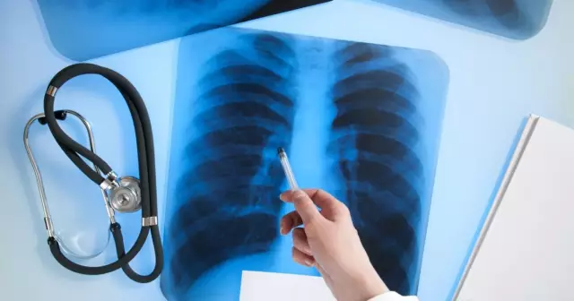
When carrying out an X-ray examination, a shadow with clear concave borders is determined on radiographs. When performing fluoroscopy in patients with atelectasis of the lung, it is possible to identify the Jacobson-Gelznecht symptom (jerky displacements of the mediastinal shadow directed towards the lesion).
Course and forecast
If the cause that led to the development of atelectasis persists, the disease lasts for a long time. When the inflammatory process joins, it gives an impetus to the development of pneumonia. If the site of atelectasis of the lung is preserved for more than six months, pneumosclerosis occurs, and then chronic pneumonia.
Atelectasis: treatment
Therapy of atelectasis of the lung should first of all be aimed at eliminating the cause of its development. With obstructive atelectasis, they resort to bronchoscopy, during which a foreign body or caseous masses are removed from the lumen of the bronchus. After restoration of the patency of the adducting bronchus, atelectasis is resolved in a short time.

Treatment of atelectasis of reflex genesis or associated with blockage of the lumen of the bronchi with mucus consists in the use of methods that accelerate the cleansing of the bronchi (stimulation of cough, massage, postural drainage).
Compression atelectasis, in cases where it is caused by a tumor or enlarged lymph nodes, requires surgical treatment.
In severe cases of atelectasis, treatment of the disease often begins with the transfer of the patient to controlled artificial ventilation.
Atelectasis in newborns
or underdevelopment of the respiratory center, underdevelopment of the lung and bronchial tissue (The main reasons for not expanding the area of lung tissue and the occurrence of atelectasis in newborns are a decrease in excitability more often in premature babies) and obstruction of the airways with mucus, blood or amniotic fluid.
The clinical picture of atelectasis in newborns is manifested by shortness of breath, tachycardia, cyanosis.
In most cases, areas of non-straightened lung tissue straighten on their own during the first week of a child's life. The prognosis of the disease deteriorates significantly with the addition of edematous-hemorrhagic syndrome, infectious and inflammatory processes or the formation of hyaline membranes.
Treatment of atelectasis in newborns consists of oxygen therapy or, in especially severe cases, artificial ventilation. Infusion therapy with glucose-alkaline mixtures is carried out, cardiovascular drugs and vitamins are prescribed.
YouTube video related to the article:
The information is generalized and provided for informational purposes only. At the first sign of illness, see your doctor. Self-medication is hazardous to health!


