- Author Rachel Wainwright [email protected].
- Public 2023-12-15 07:39.
- Last modified 2025-11-02 20:14.
Mitral valve prolapse

The mitral valve is represented by the anterior and posterior connective tissue valves, which have the appearance of flat leaves. The leaflets are attached to the papillary muscles by strong chords (threads), and the papillary muscles are attached to the bottom of the left ventricle of the heart. During the relaxation phase (diastole), the valve leaflets bend down. Blood from the left atrium moves freely into the left ventricle. In the systolic phase, the valve leaflets rise, closing the entrance to the left atrium.
Mitral valve prolapse is the sagging of the leaflets (one or both) into the atrial cavity. For the first time this phenomenon was described at the time of the appearance of the method of echocardiography (in the 60s). This pathology can be diagnosed at any age, but most often it is detected in children over seven years old.
Pathogenesis and etiology of mitral valve prolapse
Depending on the origin, mitral valve prolapse is divided into idiopathic (primary) and secondary. Primary prolapse occurs with connective tissue dysplasia. Dysplasia of connective tissue leads to a change in the structure of the papillary muscles and valve, impaired distribution, improper attachment, shortening or lengthening of the chords, and the appearance of additional chords.
Secondary mitral valve prolapse usually accompanies and complicates hereditary syndromes (congenital contracture arachnodactyly, elastic pseudoxanthoma, Ehlers-Danlo-Chernogubov syndrome, Marfin's syndrome), as well as endocrine pathologies, rheumatic diseases, heart defects. Secondary valve prolapse can develop with acquired myxomatosis, inflammatory damage to valve structures, and valve-ventricular imbalance.
Mitral valve prolapse can develop due to dysfunction of the autonomic nervous system, metabolic disorders and deficiency of trace elements, in particular potassium and magnesium ions
Divergence or loose closure of the valve leaflets is accompanied by the appearance of systolic murmur of varying intensity. In this case, mesosystolic clicks arising from excessive tension of the chords are recorded auscultatory.
Depending on the magnitude of the protrusion of the valve leaflets, the following degrees of mitral valve prolapse are distinguished:
- first degree (from 2 to 3mm);
- second degree (from 3 to 6mm);
- third degree (6 to 9 mm);
- the fourth degree of mitral valve prolapse (more than 9 mm).
The course of this disease is usually benign, long-term, favorable. The dysfunction of the valve apparatus usually progresses slowly. In some patients, the condition remains stable throughout their lives, while in other patients, this valve pathology may decrease or disappear with age.
Mitral valve prolapse symptoms
Symptoms of mitral valve prolapse depend on the severity of autonomic shifts and connective tissue dysplasia. Patients with this disease most often complain of increased fatigue, palpitations, dizziness, headaches, decreased physical performance, interruptions in the work of the heart, psychoemotional lability, irritability, increased excitability, pain in the heart, anxiety, hypochondriacal and depressive reactions.
This disease is characterized by various manifestations of connective tissue dysplasia: reduced body weight, increased skin elasticity, joint hypermobility, scoliosis, chest deformity, flat feet, myopia, pterygoid scapula. You can also find nephroptosis, sandal fissure, a peculiar structure of the gallbladder and auricles, hyperterolism of the nipples and eyes. Often, with valve prolapse, changes in blood pressure and heart rate are observed.
Diagnosis and treatment of mitral valve prolapse

Instrumental and clinical methods are used to diagnose valve prolapse. Anamnesis data, complaints, X-ray and ECG results, manifestations of connective tissue dysplasia contribute to the diagnosis. Diagnostic methods make it possible to identify this pathology and differentiate it from acquired or congenital mitral valve insufficiency, other variants of cardiac anomalies or dysfunction of the valve apparatus. According to the results of echocardiography, it is possible to correctly assess the identified cardiac changes.
The tactics of treating mitral valve prolapse depends on the degree of valve leaflet prolapse and the volume of regurgitation, as well as the nature of cardiovascular and psychoemotional disorders. With this disease, it is imperative to pay attention to sufficient (prolonged) sleep. The question of playing sports is usually considered by the attending physician after assessing the indicators of physical fitness. Patients with prolapse without significant regurgitation can lead an active lifestyle without any restrictions.
Herbal medicine is an important component in the treatment of valve prolapse. This type of treatment consists in taking sedatives (sedatives) based on motherwort, valerian, wild rosemary, hawthorn, St. John's wort, and sage. Drug therapy is aimed at symptomatic treatment of the manifestations of the disease.
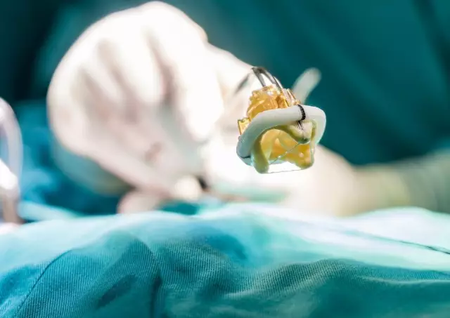
Suturing of the affected mitral valve (valvuloplasty) is performed with severe regurgitation and circulatory failure. If valvuloplasty is ineffective, the affected valve is replaced with an artificial analogue.
Complications of mitral valve prolapse
Insufficiency of the mitral valve is a fairly common complication of rheumatic heart disease. Incomplete closure of the valve leaflets and their anatomical defect contribute to a significant return of blood to the atrium. The patient is worried about shortness of breath, weakness, cough. With the development of such a complication, prosthetics of the affected valve is indicated.
Complications of mitral valve prolapse can manifest as attacks of arrhythmia and angina pectoris. These complications can be accompanied by an abnormal heart rhythm, dizziness, fainting.
YouTube video related to the article:
The information is generalized and provided for informational purposes only. At the first sign of illness, see your doctor. Self-medication is hazardous to health!


